What Actually Works for Vagus Nerve Stimulation
Vagus nerve stimulation can be achieved through FDA-approved surgical implants, transcutaneous electrical devices targeting the ear or neck, and accessible behavioral practices like slow-paced breathing and targeted cold exposure. While some social media trends exaggerate the benefits of generic techniques, clinical evidence strongly supports specific neurostimulation parameters, resonance breathing, and gut microbiome modulation for improving stress resilience and autonomic balance. The most effective methods directly target the superficial branches of the nerve or leverage the body's natural respiratory and reflex pathways to shift the nervous system into a parasympathetic state.
The Anatomy and Physiology of the Wandering Nerve
To understand how external stimuli can influence internal organ function, it is essential to first understand the unique and expansive geography of the vagus nerve. Known medically as the tenth cranial nerve (CN X), the word vagus is Latin for "wandering," a name that perfectly encapsulates its sprawling trajectory through the human body 113. It is the longest nerve of the autonomic nervous system, serving as the primary biological conduit for the parasympathetic nervous system, which governs the body's "rest and digest" physiological state 145.
The vagus nerve is not a single structure but rather a paired set of nerves - the left and right vagus - that originate in the medulla oblongata of the brainstem 112. The cellular origins of these nerves lie in specific brainstem nuclei, primarily the nucleus ambiguus and the dorsal motor nucleus of the vagus 112. Upon leaving the brainstem, the nerve fibers exit the skull through the jugular foramen, passing into the carotid sheath where they travel inferiorly alongside the internal jugular vein and the common carotid artery 123. From the neck, the branches diverge to innervate the chest and the abdomen, reaching the heart, lungs, esophagus, stomach, and the intestinal tract all the way down to the splenic flexure of the colon 13.
Crucially, the vagus nerve functions as a bidirectional communication highway. It is considered a mixed nerve, containing both motor (efferent) and sensory (afferent) fibers. However, the distribution is highly asymmetrical: approximately 80% to 90% of the fibers are afferent, meaning their primary job is to collect sensory information regarding the state of the internal organs and relay it upward to the central nervous system 149. Only the remaining 10% to 20% are efferent fibers, which carry top-down motor commands from the brain to regulate organ function, such as slowing the heart rate, constricting the pupils, and stimulating gastric acid production 4349.
Because the main trunk of the vagus nerve is buried deep within the neck and torso, directly accessing it requires understanding its superficial branches.
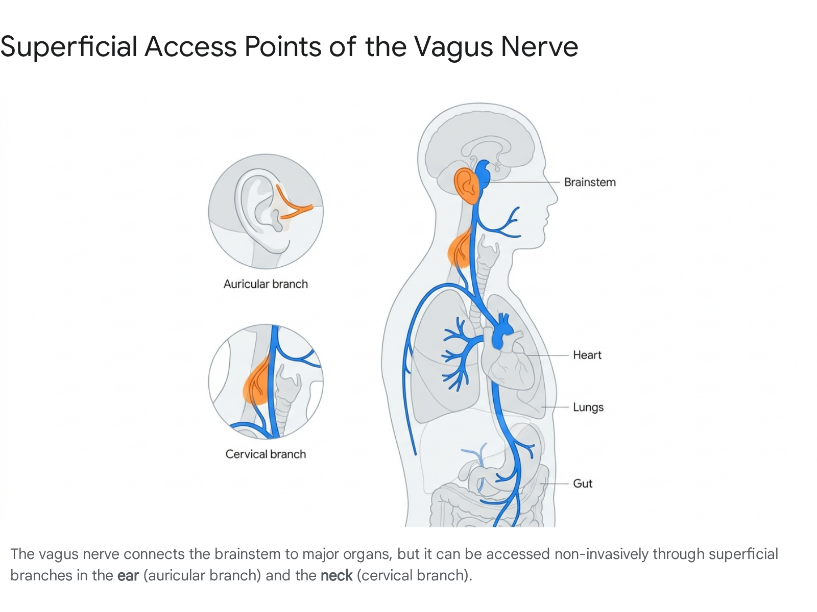
The nerve features two significant ganglia (masses of nerve tissue) near the skull base: the superior and inferior ganglia 32. The superior ganglion gives rise to the auricular branch (often referred to as Arnold's nerve), which provides sensory innervation to the posterior part of the external auditory canal and specific regions of the outer ear, such as the cymba conchae and the tragus 23511. This anatomical quirk - a cranial nerve surfacing at the outer ear - is what makes non-invasive auricular stimulation possible 56.
The functional asymmetry of the left and right vagus nerves is also of vital clinical importance. While both branches govern autonomic homeostasis, the right vagus nerve predominantly innervates the sinoatrial (SA) node - the heart's natural pacemaker - thereby having a more direct influence on modulating heart rate 1. The left vagus nerve, conversely, exerts more influence over atrioventricular conduction 1. This distinct cardiac wiring dictates exactly how and where clinical interventions are applied to maximize therapeutic benefit while minimizing cardiovascular risk.
Measuring the Invisible: Vagal Tone and Heart Rate Variability
When clinicians and researchers discuss optimizing the vagus nerve, they frequently refer to "vagal tone." This concept describes the baseline activity of the vagus nerve and its capacity to efficiently shift the body from a state of sympathetic arousal (stress) back to a state of parasympathetic calm (recovery) 1113. Because the nerve itself cannot be directly observed without invasive surgery, scientists rely on a sophisticated cardiac proxy to measure vagal tone: Heart Rate Variability (HRV) 1314.
The human heart does not beat with the perfect, rigid consistency of a metronome. Instead, the intervals between successive heartbeats fluctuate by hundreds of milliseconds 1314. This beat-to-beat variation represents a continuous, dynamic tug-of-war between the sympathetic nervous system, which attempts to accelerate the heart, and the parasympathetic nervous system (driven by the vagus nerve), which attempts to slow it down 14. A healthy, adaptable nervous system exhibits high HRV, indicating that the vagus nerve is actively applying the brakes and allowing the heart to quickly respond to environmental demands 1314. Conversely, chronically stressed individuals exhibit low HRV, signifying a system stuck in sympathetic overdrive with diminished vagal influence 14.
The Metrics of Vagal Modulation
To accurately quantify the vagus nerve's contribution to this variability, researchers analyze electrocardiogram (ECG) data using specific time-domain and frequency-domain parameters 789.
In the time domain, the most widely recognized marker of vagal tone is RMSSD (Root Mean Square of Successive Differences) 1389. RMSSD calculates the variance between adjacent heartbeats and is highly sensitive to the high-frequency beat-to-beat alterations driven by the vagus nerve, remaining relatively uninfluenced by slower sympathetic changes 139. Another common time-domain measure is SDNN (Standard Deviation of NN intervals), which reflects overall autonomic balance rather than isolated parasympathetic activity 89.
In the frequency domain, HRV is segmented into spectral bands. The High-Frequency (HF) band, typically defined as frequencies between 0.15 and 0.40 Hz, is heavily mediated by vagal activity and is closely aligned with the respiratory cycle 89. The Low-Frequency (LF) band (0.04 to 0.15 Hz) represents a complex mixture of both sympathetic and parasympathetic influences, alongside thermoregulation and baroreflex activity 9. Researchers often use the LF/HF ratio to evaluate the balance between sympathetic and parasympathetic tone, though this metric remains heavily debated in the literature 810.
Obtaining accurate vagal tone measurements is technically demanding. Standard consumer smartwatches may underestimate the complex frequency content of the ECG signal, particularly the sharp, Dirac-shaped R-peaks of the heartbeat. Leading researchers suggest that professional HRV analysis requires high-resolution recordings (e.g., 8,000 Hz sampling rates) and specialized filtering to capture the rapid fluctuations caused by vagal activity without introducing artificial digitization noise 19. Consequently, while consumer wearables provide useful trend data, true clinical assessment of vagal tone relies on rigorous, controlled ECG measurements 719.
Table: Key Heart Rate Variability Metrics for Vagal Tone
| Metric Category | Specific Parameter | Physiological Origin & Significance | Clinical Interpretation |
|---|---|---|---|
| Time-Domain | RMSSD | Measures successive heartbeat differences. Highly sensitive to vagal input. 139 | The primary proxy for parasympathetic (vagal) tone. Higher values indicate resilience. 138 |
| Time-Domain | SDNN | Measures overall variance across all cyclic components during the recording period. 89 | Reflects broad autonomic balance (both sympathetic and parasympathetic). 8 |
| Frequency-Domain | High-Frequency (HF) | Ranges from 0.15 to 0.40 Hz. Corresponds to the respiratory cycle. 89 | Directly reflects vagal tone and respiratory sinus arrhythmia. 89 |
| Frequency-Domain | Low-Frequency (LF) | Ranges from 0.04 to 0.15 Hz. Represents baroreflex, hormones, and thermoregulation. 9 | A mixed marker reflecting both sympathetic and parasympathetic activity. 9 |
Bioelectronic Medicine: The Evolution of Electrical Stimulation
The most rigorously validated method for stimulating the vagus nerve lies in the realm of bioelectronic medicine, utilizing precise electrical impulses to depolarize nerve fibers and trigger systemic physiological cascades. This approach has evolved over three decades from highly invasive surgical procedures to sophisticated, non-invasive wearable technologies 411.
Surgically Implanted Vagus Nerve Stimulation (iVNS)
Invasive Vagus Nerve Stimulation (iVNS) represents the foundational clinical application of this technology. First utilized in the 1980s by American neurologist James Corning, the modern era of iVNS began when the U.S. Food and Drug Administration (FDA) approved surgically implanted devices for the treatment of drug-resistant epilepsy in 1997 414. Subsequent approvals followed for treatment-resistant depression in 2005, and more recently, for stroke rehabilitation 1213.
The surgical procedure involves implanting a titanium-encased pulse generator (similar to a cardiac pacemaker) beneath the skin in the upper left chest 1214. A lead wire is tunneled subcutaneously up the neck and coiled directly around the left cervical vagus nerve 1213. The left nerve is almost exclusively chosen for these implants because the right vagus nerve more densely innervates the sinoatrial node; stimulating the right side carries a higher risk of inducing profound bradycardia or cardiac arrhythmias 12. The device is programmed by a physician to deliver intermittent bursts of electricity - typically operating at frequencies of 20 to 30 pulses per second (Hz) for several seconds, followed by several minutes of rest 13.
Longitudinal data underscores the efficacy of iVNS. For patients with drug-resistant epilepsy, a review of over 4,000 registry patients demonstrated a 46% reduction in seizure frequency after three months, with benefits compounding to a 75% reduction after ten years of continuous therapy 415. In psychiatry, long-term studies reveal that over 50% of patients with chronic, treatment-refractory depression experience a clinically significant reduction in symptoms following iVNS implantation 4.
Furthermore, iVNS has opened entirely new therapeutic frontiers. In 2025, the FDA approved the SetPoint System, a pioneering implantable device designed specifically to treat rheumatoid arthritis 1617. This application emerged from decades of research by Dr. Kevin J. Tracey, who identified the "cholinergic anti-inflammatory pathway" - a mechanism by which efferent vagal signals instruct the spleen to halt the production of inflammatory cytokines like TNF-alpha 416.
However, iVNS is not without significant drawbacks. Surgical implantation is expensive, typically costing tens of thousands of dollars, and it subjects the patient to operative risks 11. A 25-year longitudinal registry study of 247 patients found an 8.6% surgical complication rate, which included postoperative hematomas (1.9%), infections (2.6%), and vocal cord palsy (1.4%) 18. Additionally, the hardware itself can fail, with lead fractures occurring in approximately 3.0% of cases 18. When the device is actively stimulating, patients frequently report side effects such as voice alteration, coughing, throat pain, and shortness of breath 1319. Because of these risks, researchers began aggressively pursuing non-invasive alternatives.
Transcutaneous Vagus Nerve Stimulation (nVNS)
To harness the therapeutic power of the vagus nerve without the risks of surgery, medical engineers developed transcutaneous (through the skin) vagus nerve stimulation (nVNS). These devices deliver mild electrical microcurrents to the superficial branches of the vagus nerve, utilizing specific anatomical access points on the neck and the ear 5629. Animal studies utilizing c-Fos brain mapping have demonstrated that non-invasive stimulation yields convergent patterns of neural activation in the brainstem compared to invasive implants, cementing the biological plausibility of the transcutaneous approach 3020.
Transcutaneous Cervical VNS (tcVNS)
Cervical nVNS targets the main trunk of the vagus nerve as it travels through the carotid sheath in the neck 2921. These handheld devices, such as the FDA-cleared gammaCore and the consumer-equivalent Truvaga Plus, are positioned against the side of the neck and deliver proprietary electrical waveforms (often at a 25 Hz frequency) 2933. The electrical pulse penetrates the skin to depolarize the underlying nerve fibers.
Clinical research into tcVNS has been highly successful in the realm of neurology, leading to regulatory approvals for the acute and preventative treatment of cluster headaches and migraines 1229. Beyond pain management, studies suggest that tcVNS can modulate autonomic tone. In laboratory settings, applying tcVNS during acute mental and physiological stressors has been shown to counteract stress-induced sympathetic arousal, significantly altering cardiac timing metrics like the Pre-Ejection Period (PEP) and reducing pulse pressure 21.
Transcutaneous Auricular VNS (taVNS)
Auricular stimulation (taVNS) has become one of the most intensely researched areas of neuromodulation. The rationale relies on the unique anatomy of the outer ear, which is the only cutaneous region of the human body innervated by the vagus nerve, specifically via the auricular branch 56. Researchers have identified the cymba conchae and the inner tragus as the optimal anatomical targets; stimulating these specific cartilaginous indentations elicits higher vagus somatosensory evoked potentials (VSEPs) in the brainstem compared to other areas of the ear 62022.
Devices designed for taVNS typically resemble traditional earbuds or ear clips 1421. When powered on, they deliver microcurrents (often using 200 μs pulses at frequencies ranging from 10 to 30 Hz) that travel along the auricular nerve directly into the nucleus tractus solitarius in the brainstem 102023. A massive influx of clinical trials between 2020 and 2025 has explored taVNS across a wide spectrum of pathologies.
Systematic reviews covering over 100 studies indicate that taVNS exerts a favorable therapeutic effect in populations suffering from epilepsy, depression, and post-stroke motor deficits 3024. In cardiovascular research, taVNS application has been shown to reduce both systolic and diastolic blood pressure in hypertensive individuals while simultaneously increasing resting RMSSD (vagal tone) 1022. The therapy has also demonstrated promise in psychiatric settings; a 2024 trial evaluating taVNS combined with repetitive transcranial magnetic stimulation (rTMS) for moderate-to-severe depression yielded an impressive 85% overall response rate, significantly outperforming monotherapy 25.
However, the efficacy of taVNS remains debated in certain complex, multi-systemic conditions. For instance, while taVNS has been heavily marketed as a potential treatment for Long COVID (post-COVID-19 condition) to combat neuroinflammation and dysautonomia, rigorous evidence is lacking. A 2025 PRISMA-compliant systematic review of taVNS in Long COVID patients found the certainty of evidence to be "very low" 23. In one double-blinded randomized controlled trial, the sham (placebo) stimulation paradoxically produced greater improvements in fatigue than the active taVNS device, despite the active device successfully raising HRV 23. This highlights a persistent challenge in nVNS research: the difficulty of designing a perfect sham protocol, as patients can often feel the electrical tingling of the active device 24.
The Expanding VNS Market and Device Landscape
Driven by the shift from surgical implants to non-invasive therapies, the global vagus nerve stimulation market has experienced explosive growth. Valued at approximately $575 million in 2025, the market is projected to reach nearly $1 billion by the early 2030s, expanding at a compound annual growth rate (CAGR) of roughly 6% to 9% 1517. North America currently dominates this sector, commanding over 50% of global market share due to advanced healthcare infrastructure, early adoption of neuromodulation, and the strong presence of major manufacturers like LivaNova and electroCore 151738. However, the Asia-Pacific region is experiencing the fastest growth, propelled by rising healthcare expenditures and a growing acceptance of non-invasive mental health interventions 173826.
The commercialization of nVNS has resulted in a crowded market of consumer wellness devices, which range widely in price, underlying technology, and clinical validation. It is crucial to distinguish between true electrical VNS (which directly depolarizes the nerve) and vibrational/acoustic wearables, which aim to activate the parasympathetic system indirectly via mechanoreceptors or bone conduction 33.
Table: Comparison of Prominent Non-Invasive VNS and Modulatory Devices
| Device Name | Technology Type | Target Location | Estimated Cost (2025) | Clinical Validation & Notes |
|---|---|---|---|---|
| gammaCore | Electrical tcVNS | Cervical (Neck) | Prescription pricing | High: FDA-cleared for cluster headache and migraine. Strong clinical data. 2927 |
| Truvaga Plus | Electrical tcVNS | Cervical (Neck) | ~$499 | Moderate-High: Consumer equivalent of gammaCore utilizing similar 25 Hz parameters. 293341 |
| Nuropod (Nurosym) | Electrical taVNS | Auricular (Tragus) | ~$900 | High: Extensive clinical trials with major universities; FDA Non-Significant Risk designation. 33 |
| Sona | Electrical taVNS | Auricular (Ear) | ~£695 | Moderate: Utilizes AI closed-loop systems to adapt stimulation based on real-time HRV feedback. 1129 |
| Pulsetto | Electrical tcVNS | Cervical (Neck) | ~$278 | Moderate: Bilateral neck stimulation. Very affordable entry point but requires app subscription for full features. 29334142 |
| Apollo Wearable | Vibrational Haptics | Wrist / Ankle | ~$300 | Indirect: Uses low-frequency sound waves/vibration. Pilot studies show HRV benefits, but it does not electrically stimulate the vagus. 1433 |
| Sensate | Infrasonic Resonance | Sternum (Chest) | ~$299 | Indirect: Bone conduction vibration on the chest wall. Popular for relaxation, lacks robust clinical trials. 1441 |
Thermal Interventions: The Science of Cold Exposure
In recent years, the wellness industry and social media platforms have aggressively promoted cold exposure as a mechanism for instantly "resetting" the vagus nerve. Viral videos on platforms like TikTok claim that placing a bag of frozen peas on the chest or taking a freezing ice bath will cure insomnia and eliminate anxiety by triggering parasympathetic pathways 4282930. While there is a genuine physiological basis for cold-induced vagal activation, the biological mechanisms are far more nuanced - and anatomically specific - than popular narratives suggest.
The primary mechanism linking cold exposure to the vagus nerve is the mammalian dive reflex, also known in clinical literature as the trigeminocardiac or trigeminovagal reflex 4631. This is an ancient, evolutionary conservation response observed across many mammalian species. When the face is submerged in cold water, thermal receptors in the skin activate the trigeminal nerve (Cranial Nerve V), which is the primary sensory nerve of the face 46. The trigeminal nerve sends an immediate signal to the brainstem, which in turn commands the efferent fibers of the vagus nerve (Cranial Nerve X) to drastically slow the heart rate (bradycardia) and constrict peripheral blood vessels 463132. This rapid parasympathetic shift is designed to conserve oxygen during underwater submersion.
Research confirms that you do not need to hold your breath underwater to trigger this vagal response. Studies demonstrate that simply applying cold stimulus to specific locations - namely the forehead, cheeks, and the lateral neck - reliably increases RMSSD and slows the heart rate 284933. In a pivotal 2018 randomized controlled trial, researchers utilized precisely calibrated thermodes to apply cold for 16-second intervals to subjects' necks, cheeks, and forearms 2849. The data revealed that heart rate decreased and HRV increased only when the cold was applied to the neck and cheeks, completely failing to produce an autonomic shift when applied to the forearm 2849. This spatial specificity confirms that the relaxing effect is driven by the anatomical proximity of the trigeminal and vagal sensory receptors, not merely the subjective sensation of being cold 2849.
Therefore, placing a bag of frozen peas on the sternum (chest) is physiologically misaligned with the neural architecture required to trigger the dive reflex 49. While the chest application may feel subjectively soothing, it lacks the direct cranial nerve pathways necessary to reliably alter vagal tone.
The effects of full-body cold-water immersion (such as ice baths or cold plunging) are even more complex. Full-body cold exposure is a nonselective, intense physiological stressor 3046. Entering freezing water instantly triggers a massive sympathetic nervous system spike - often referred to as "cold shock" - characterized by hyperventilation, a surge of adrenaline, and a rapid increase in heart rate 3046. The parasympathetic, vagal response only becomes dominant later, primarily as the body attempts to regain homeostasis and rewarm itself 3031. While regular cold-water swimmers do exhibit higher baseline vagal tone in the long term, using a sudden ice bath as a precision tool for immediate stress relief is counterproductive, as the initial shock inherently triggers a fight-or-flight response 304649.
For individuals seeking a targeted, manageable parasympathetic shift without the intense metabolic demands of an ice bath, clinical evidence points toward gentle, sustained cooling of the lateral neck or splashing cold water directly onto the face 464951.
Respiratory and Mechanical Modulation: Breathwork, Yoga, and Vibration
While bioelectronics and cold thermogenesis are highly effective, the most universally accessible method for stimulating the vagus nerve is entirely free and internally generated: controlling the mechanics of respiration.
The heart and the lungs are inextricably linked by a phenomenon known as Respiratory Sinus Arrhythmia (RSA). This is a naturally occurring physiological mechanism where the heart rate synchronizes with the breathing cycle 834. During inhalation, the lungs expand, which mildly inhibits vagal outflow; this allows the sympathetic nervous system to momentarily take control, increasing the heart rate to circulate oxygenated blood efficiently 8. Conversely, during exhalation, vagal efferent fibers are stimulated. The vagus nerve releases the neurotransmitter acetylcholine directly into the sinoatrial node, rapidly applying the brakes and slowing the heart rate down 1138.
By consciously altering the pace and depth of breathing, individuals can hijack this autonomic reflex to manually increase vagal tone. Scientific consensus indicates that "resonance frequency breathing" - a deliberate pattern of slowing respiration to approximately 4.5 to 7 breaths per minute - maximizes the amplitude of RSA 13834. By extending the exhalation phase to be longer than the inhalation phase, practitioners prolong the period of vagal activation, leading to immediate, measurable improvements in HRV and a profound shift toward parasympathetic dominance 1334.
The Clinical Efficacy of Pranayama and Yoga
These physiological mechanics provide the neurobiological explanation for why traditional Eastern practices, such as yoga and Pranayama (yogic breathing), are highly effective at mitigating stress and clinical depression 535. Extensive literature reviews confirm that structured Pranayama practice broadens vagal modulation, decreases cortisol production, and enhances the release of neurotrophic factors in the brain 736.
Specific breath retention techniques, such as Kumbhak, rely on a combination of physiological triggers. Holding the breath deliberately alters intrathoracic pressure, which stimulates baroreceptors in the chest and neck, while the resulting mild hypoxia and increased carbon dioxide tolerance paradoxically improve tissue oxygenation via the Bohr effect 37. These pressure and chemical changes serve as potent activators of the vagus nerve 37.
The psychiatric benefits of these practices are well-documented. Studies utilizing functional MRI have demonstrated that a four-week regimen of Bhastrika Pranayama significantly reduces clinical anxiety by modulating connectivity between the prefrontal cortex and the anterior insula - brain regions heavily involved in emotional processing 5. Similarly, large-scale studies conducted by researchers at Harvard Medical School evaluating specific practices taught by yogi Sadhguru (such as Isha Kriya and Upa Yoga) revealed profound reductions in psychological distress; regular practitioners saw anxiety levels drop by up to 33% and symptoms of depression decrease by 50% within just a few weeks 38.
Mechanical Vibration: Chanting and Ear Massage
Because the vagus nerve features branches that innervate the vocal cords (the recurrent laryngeal nerve) and the outer ear (the auricular branch), mechanical manipulation of these areas can stimulate the nerve fibers 11.
Audible, rhythmic vocalizations act as a form of internal vibration therapy. Clinical neurophysiology studies examining the practice of loud "OM" chanting reveal that the specific vibrational sensations in the larynx and auricular structures directly stimulate vagal pathways 34. A study measuring autonomic function during five minutes of OM chanting found an immediate increase in high-frequency HRV power (parasympathetic dominance) and a corresponding deactivation of the brain's limbic and paralimbic structures, which are responsible for generating emotional stress responses 34. Importantly, control subjects performing simple "ssss" sounds did not exhibit these autonomic shifts, suggesting the specific frequency and vibration of the chant are mechanically significant 34.
Externally, practitioners can utilize ear massage to target the auricular vagus nerve. By gently applying pressure to the tragus or rubbing the cymba conchae in slow, circular motions, individuals can provide mild mechanical stimulation to the underlying nerve fibers 11. While the physiological impact of manual ear massage is undoubtedly weaker than the targeted microcurrents of a clinical taVNS device, therapists frequently employ it as an effective, immediate grounding technique to help patients regulate their nervous systems during panic attacks or trauma responses 1157.
The Microbial Vagus: The Gut-Brain Highway
Perhaps the most groundbreaking frontier in vagal research is the gut-brain axis. The vagus nerve operates as the primary physical bridge connecting the central nervous system to the enteric nervous system of the gastrointestinal tract 9395940. Because the vast majority of vagal fibers are sensory, the nerve essentially acts as a massive biological antenna, constantly monitoring the state of the gut microbiome and reporting back to the brain 94041.
The communication between gut microbes and the vagus nerve is highly sophisticated. Trillions of bacteria reside in the gut, breaking down dietary fibers and producing a variety of active metabolites, including short-chain fatty acids (SCFAs) like butyrate, as well as bile acids and neurotransmitter precursors 94142. While vagal sensory fibers do not physically cross the intestinal epithelial lining, these bacterial metabolites seep through the barrier and bind to specialized receptors on the nerve's intraganglionic laminar endings 9.
A landmark 2025 study from UCLA provided definitive proof of this causal relationship. Researchers utilized germ-free mice - animals bred entirely without a gut microbiome - and observed that their vagal nerve activity was severely diminished compared to normal mice 42. When the researchers introduced specific microbial metabolites (SCFAs and bile acids) into the intestines of these germ-free mice, they recorded direct, immediate electrical activation within the vagus nerve, which subsequently triggered neurons in the brainstem 42. This confirms that the gut microbiome dictates vagal tone.
When the gut microbiome falls into a state of imbalance - known as dysbiosis - this critical communication channel degrades. A loss of beneficial bacteria results in fewer SCFAs, impaired vagal signaling, and a consequent rise in systemic inflammation 95943. This inflammatory state can travel up the vagus nerve and breach the blood-brain barrier, contributing significantly to the pathogenesis of neurodegenerative diseases, major depressive disorder, and severe anxiety 414344.
Consequently, researchers are actively investigating how to stimulate the vagus nerve "from the bottom up" using targeted probiotics, often referred to as psychobiotics. Clinical data is highly promising. A 2025 study evaluating 86 participants found that patients suffering from major depression exhibited significantly compromised vagal function compared to healthy controls 40. However, after administering a specific multi-species probiotic (OMNi-BiOTiC) for three months, the depressed patients showed a significant increase in healthy bacteria (specifically Akkermansia muciniphila) and a corresponding, measurable improvement in morning vagal tone (via HRV), which correlated with enhanced sleep quality and reduced depressive symptoms 40. Cultivating a diverse, healthy microbiome through a fiber-rich diet and validated probiotic strains provides a sustained, long-term foundation for optimizing vagal function and mental health 943.
The Dangers of Overstimulation: The Oculocardiac Reflex
While increasing vagal tone is broadly beneficial for managing stress and inflammation, the vagus nerve is a powerful regulator of cardiovascular function. Aggressive or misguided attempts to stimulate it can lead to severe, potentially fatal medical complications.
One of the most profound examples of dangerous vagal overstimulation is the oculocardiac reflex (also known as the Aschner-Dagnini reflex) 324546. This is a physiological reflex arc initiated by applying pressure, traction, or compression directly to the eyeball or the extraocular muscles 3245. The sensory (afferent) limb of this reflex is driven by the ophthalmic branch of the trigeminal nerve, which senses the pressure on the eye and relays a distress signal to the reticular formation in the brainstem 3246. There, the signal synapses with the motor (efferent) nucleus of the vagus nerve, which immediately sends an overwhelming inhibitory signal down to the sinoatrial node of the heart 3246.
The result is a precipitous and dangerous drop in heart rate (bradycardia). In clinical settings, particularly during strabismus (crossed-eye) correction surgery in children or during maxillofacial trauma procedures, traction on the eye muscles can cause the heart rate to plummet by more than 50% 324546. In up to 10% of these surgical cases, the vagal response is so profound that it induces cardiac arrhythmias, severe hypotension, and asystole (complete cessation of the heartbeat), requiring immediate cessation of the stimulus, administration of intravenous anticholinergic drugs like atropine, and potentially cardiopulmonary resuscitation 324546.
While ocular compression has historically been documented in veterinary medicine as a crude method for aborting seizures in dogs, it is universally condemned as a health practice for humans 476869. Attempting to "hack" the vagus nerve by applying deep pressure to the eyes poses an unacceptable risk of triggering life-threatening bradycardia 193246.
Furthermore, general precautions apply to all forms of vagus nerve stimulation. Individuals with a history of cardiac conduction disease, arrhythmias, severe bradycardia, or those utilizing implanted medical devices (such as pacemakers or defibrillators) should explicitly avoid electrical nVNS devices and extreme cold plunges unless strictly monitored by a cardiologist 19. Because the vagus nerve inherently lowers blood pressure, patients prone to orthostatic hypotension or syncope (fainting) must proceed with extreme caution, as overstimulation can easily trigger a fainting episode 19.
Bottom line
The vagus nerve serves as the master controller of the body's parasympathetic recovery system, providing a vital counterbalance to chronic stress and inflammation. Clinical evidence confirms that the nerve can be effectively stimulated through advanced bioelectronic medicine - ranging from FDA-approved surgical implants for severe neurological diseases to accessible, non-invasive transcutaneous wearables that target the ear and neck. For those seeking entirely natural interventions, resonance breathing, vocal vibration, targeted facial cold exposure, and microbiome optimization via probiotics offer scientifically validated pathways to increase vagal tone. However, the cardiovascular power of the vagus nerve demands respect; consumers should avoid intense, non-selective stressors like unacclimated full-body ice baths and entirely reject dangerous physical maneuvers like ocular compression.