How the Glymphatic System Cleans Your Brain During Sleep
The glymphatic system is the brain's highly specialized, built-in waste clearance network that relies on cerebrospinal fluid to flush out neurotoxic metabolic byproducts, including amyloid-beta and tau proteins. This microscopic plumbing system operates almost exclusively during deep sleep, utilizing rhythmic blood vessel pulsations driven by chemical signals to physically pump fluid through dense neural tissue. When sleep is chronically disrupted, this mechanical washing cycle stalls, directly contributing to the accumulation of proteins that are heavily associated with Alzheimer's disease and other neurodegenerative conditions.
The Brain's Unique Waste Management Challenge
The human brain represents a profound biological paradox. Despite accounting for only about 2% of total body mass, the central nervous system consumes a disproportionate 20% of the body's energy to maintain continuous electrical signaling and synaptic transmission 1. This exceptionally high metabolic rate produces a massive, continuous output of cellular waste. The byproducts of neural activity include lactic acid, excess potassium, and stray proteins like amyloid-beta and tau, all of which become toxic to the delicate neural circuitry if allowed to accumulate 12.
In the rest of the body, the peripheral lymphatic system serves as a biological sewage network. A dense, dedicated web of lymphatic vessels collects excess interstitial fluid (the fluid dwelling between cells), cellular debris, and immune cells. This lymph fluid is filtered through lymph nodes before being returned to the bloodstream 134.
However, the brain and spinal cord are locked behind the tightly regulated blood-brain barrier. For centuries, anatomists observed that the functional tissue of the brain - the parenchyma - lacks these traditional lymphatic vessels entirely 35. This physiological reality presented a massive question for neurobiology: How does the most metabolically active organ in the body survive without a conventional waste-drainage system?
For a long time, the prevailing assumption was that the brain relied on simple, passive diffusion to clear its waste, a process that would be highly inefficient given the density of the tissue 56. The definitive answer arrived in 2012, when Danish neuroscientist Maiken Nedergaard and her research team at the University of Rochester utilized in-vivo two-photon microscopy to reveal a previously hidden, highly organized fluid transport superhighway 457.
Nedergaard coined the term "glymphatic system," a portmanteau reflecting the pathway's absolute reliance on glial cells to perform a lymphatic-like function 567.
Structural Anatomy of the Clearance Pathway
Rather than building a separate network of drainage pipes, evolution engineered the brain to co-opt the exterior architecture of its existing blood vessels. The glymphatic mechanism operates through a polarized, macroscopic system of convective fluid fluxes 14.
The clearance process follows a distinct three-step anatomical pathway:
- Periarterial Influx: Cerebrospinal fluid (CSF), the clear liquid that surrounds and protects the brain, is driven into the deep brain tissue along perivascular spaces - specifically known as Virchow-Robin spaces. These are essentially microscopic tunnels that form the outer boundary of penetrating cerebral arteries 234.
- Astrocyte Checkpoints and Fluid Mixing: The boundary between this perivascular space and the dense brain parenchyma is completely lined by the "endfeet" of astrocytes, a specialized type of star-shaped glial cell. These astrocyte endfeet are heavily studded with water channels called Aquaporin-4 (AQP4). The AQP4 channels act as selective valves, pulling clean CSF from the perivascular space directly into the brain tissue. Once inside, the CSF mixes with the interstitial fluid (ISF), collecting the accumulated metabolic waste 2356.
- Perivenous Efflux: The newly formed, waste-laden fluid mixture is swept across the tissue via convective bulk flow. It is ultimately pushed outward toward the cerebral veins, traveling along perivenous spaces. The fluid exits the brain tissue and eventually drains into the true meningeal lymphatic vessels (MLVs) - a distinct network in the membranes surrounding the brain discovered in 2015 - which carry the waste down to the deep cervical lymph nodes for systemic elimination 138.
Glymphatic vs. Peripheral Lymphatic Architecture
To fully grasp the ingenuity of the glymphatic system, it is helpful to compare its unique anatomical workarounds against the rest of the body's standard clearance infrastructure. As detailed in comparative studies, the brain's solution relies entirely on existing cellular architecture rather than dedicated vessels 2358.
| Feature | Peripheral Lymphatic System | Glymphatic System |
|---|---|---|
| Primary Location | Throughout the body (tissues, organs) | Central Nervous System (Brain and Spinal Cord) |
| Structural Conduits | Dedicated, distinct lymphatic vessels equipped with one-way valves | "Borrowed" perivascular spaces surrounding existing arteries and veins |
| Fluid Handled | Lymph (plasma, white blood cells, cellular debris) | Cerebrospinal fluid (CSF) mixing with Interstitial fluid (ISF) |
| Cellular Dependency | Endothelial cells lining the walls of the lymph vessels | Astroglial cells (Astrocytes) and their Aquaporin-4 (AQP4) water channels |
| Peak Activity Period | Continuous, scaled to physical movement and local metabolism | Highly state-dependent; heavily restricted during wakefulness, peaking in deep sleep |
| System Integration | Drains directly into the subclavian veins near the heart | Drains into meningeal lymphatics, then to deep cervical lymph nodes |
The lack of structural valves in the glymphatic system - unlike the one-way gates found in peripheral lymphatics - means the brain must rely on other biological forces to propel the fluid in a single direction. This propulsion is achieved through a mixture of arterial pulsation and diffusion, creating a net directionality toward the venous system 388.
Direct Visualization in the Living Human Brain
For over a decade following its discovery in 2012, the glymphatic system was a subject of intense scientific debate. While fluorescent tracers and real-time imaging of living mice strongly supported the hypothesis, observing this microscopic, dynamic fluid movement in living humans was technologically daunting. Many scientists remained skeptical, questioning whether the fluid dynamics observed in the rodent brain translated accurately to the massive, highly folded human cortex 7109.
That skepticism was fundamentally dismantled in October 2024 with a landmark clinical study published in the Proceedings of the National Academy of Sciences (PNAS). Researchers at Oregon Health & Science University (OHSU), led by neurologist Dr. Juan Piantino and Dr. Erin Yamamoto, provided the first definitive imaging evidence of the glymphatic system actively operating in the living human brain 7101011.
The OHSU team capitalized on a unique clinical scenario. Five patients undergoing neurosurgery for brain tumor removal consented to have an inert, gadolinium-based contrast agent injected into their cerebrospinal fluid via a lumbar drain. Following the surgical procedure, the patients underwent serial magnetic resonance imaging (MRI) using a specific technique called fluid attenuated inversion recovery (FLAIR) at 12, 24, and 48-hour intervals 7101014.
The sequential imaging results were unequivocal. The tracking revealed that the cerebrospinal fluid did not simply diffuse randomly through the human brain tissue like water soaking evenly into a sponge. Instead, the gadolinium contrast moved purposefully along clearly defined anatomical channels - the perivascular spaces. The imaging showed centripetal flow, moving from the subarachnoid space deep into the brain parenchyma, precisely mirroring the glymphatic pathway defined in rodent models 79101112. This proven structural confirmation established that the human brain possesses an identical, functioning fluid-conduit network 1014.
Why the Glymphatic System Demands Sleep
The most profound characteristic of the glymphatic system is its severe state-dependency. The network is functionally suppressed during wakefulness and becomes highly active only when the brain transitions into sleep.
When an individual is awake, the brain prioritizes sensory processing, motor control, and continuous cognition. To facilitate rapid electrical signaling, the interstitial space (the microscopic gaps between brain cells) is kept relatively tight and compact. Research utilizing advanced photo-imaging in mice demonstrates a 90% reduction in glymphatic clearance during wakefulness compared to the sleeping state 13.
The transition to sleep triggers a dramatic neuro-architectural shift. As the brain switches from active information processing to a biological maintenance mode, neuronal activity drops, and the brain cells physically shrink. This cellular contraction expands the volume of the interstitial space by up to 60% 131718. This physical expansion drastically lowers the tissue resistance, effectively opening the floodgates for CSF to flow freely and wash away the day's accumulated metabolic toxins 1718.
The 50-Second Norepinephrine Pump: A 2025 Breakthrough
While the 60% expansion of the interstitial space explained where the fluid went, researchers still lacked a complete understanding of the mechanical engine driving the flow. The ambient arterial pulse of the heartbeat pushes fluid into the entryways of the perivascular space, but it is not forceful enough to drive the fluid deep through the dense parenchymal tissue at the speeds observed during sleep 8.
In January 2025, a groundbreaking study published in the journal Cell by Dr. Natalie Hauglund and her colleagues at the University of Copenhagen and the University of Oxford completely rewrote our understanding of this mechanism 14151617. By utilizing optogenetics and simultaneous monitoring technologies to observe fluid dynamics in naturally sleeping mice, the team identified a strictly synchronized mechanical pump driven by neurochemical oscillations.
They discovered that during deep, non-rapid eye movement (NREM) sleep, a region of the brainstem called the locus coeruleus releases tiny, regular pulses of the neurotransmitter norepinephrine approximately once every 50 seconds 15161723.
Norepinephrine typically acts as an arousal and stress chemical. During waking hours, high, continuous levels of norepinephrine keep the brain's blood vessels slightly constricted and the perivascular spaces closed 623. However, during deep sleep, the overall continuous tone drops entirely, and these ultra-slow, 50-second oscillatory waves take over.
Each wave of norepinephrine triggers the brain's blood vessels to gently constrict and then relax. This synchronized phenomenon, known as "slow vasomotion," creates a rhythmic, peristaltic pumping effect. The vessels act as a thermodynamic pump, mechanically forcing cerebrospinal fluid through the brain tissue in massive, sweeping waves 81415162318.
The Ambien Paradox: Behavioral vs. Physiological Sleep
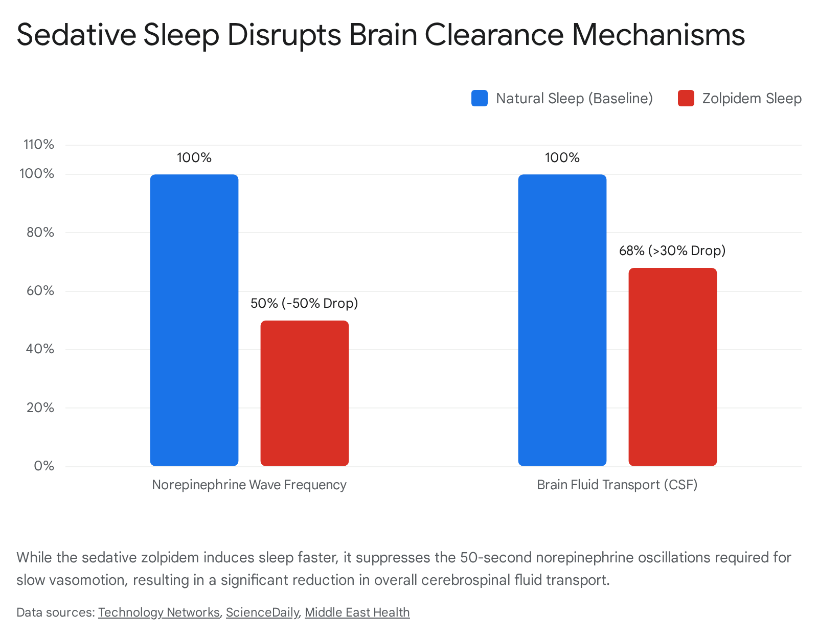
The discovery of the 50-second norepinephrine pump carries massive implications for sleep medicine, particularly regarding how we view pharmaceutical sleep aids. Hauglund's 2025 study explicitly investigated the physiological effects of zolpidem (commonly prescribed under the brand name Ambien), a widely used sedative-hypnotic drug 151618.
The researchers found that while zolpidem successfully induced behavioral sleep - allowing the subjects to fall asleep faster - it severely disrupted the underlying restorative physiology. In subjects treated with zolpidem, the critical norepinephrine waves were dampened and reduced in frequency by 50%. As a direct mechanical consequence of this chemical disruption, the pulsing slow vasomotion of the blood vessels was impaired, leading to a catastrophic drop in brain fluid transport and waste clearance of more than 30% compared to natural sleep 151618.
This data highlights a vital clinical insight: not all unconscious states are biologically equivalent. Artificial sleep induced by sedatives may quiet the conscious mind, but it can fail to activate the brain's mechanical washing cycle, leaving neurotoxic waste behind to accumulate 141516.
How Different Sleep Stages Impact Clearance Efficiency
Because the glymphatic system relies heavily on specific neurochemical states, slow-wave electrical activity, and vascular rhythms, its efficiency fluctuates dramatically depending on the exact stage of sleep a person is in.
| Sleep Stage | Glymphatic Functionality | Key Physiological Drivers and Mechanisms |
|---|---|---|
| Wakefulness | Minimal (Basal) | High, continuous norepinephrine tone keeps perivascular spaces closed. High neuronal firing restricts the interstitial space volume. Total fluid clearance drops by approximately 90% compared to sleep 21317. |
| Light NREM (N1 & N2) | Moderate | Brain transitioning. Sleep spindles generated in N2 help regulate arousal. Glymphatic clearance begins to accelerate as fluid resistance drops, but lacks maximum mechanical pumping force 1825. |
| Deep NREM (N3) | Maximum (Optimal) | Often called "slow-wave sleep," defined by high-amplitude brainwaves (>75 μV) and the lowest overall arousal. The 50-second norepinephrine vasomotion rhythm acts as a powerful mechanical pump. Interstitial space expands maximally 2131425. |
| REM (Dreaming) | Significantly Reduced | High cholinergic tone induces sustained vasodilation, restricting the necessary pulsatory motions. Cerebral electrical activity closely resembles wakefulness, largely stalling brain-wide glymphatic efficiency 132519. |
Interestingly, the Hauglund study noted that brief, subtle awakenings known as "micro-arousals" - which sleepers are entirely unaware of - actually correlate positively with glymphatic flow 23. While micro-arousals are traditionally viewed as a sign of fragmented, poor-quality sleep, they appear to occur in tandem with the mechanical flushing process during NREM phases, suggesting they are a normal feature of the physiological wash cycle rather than a disruption 23.
Conversely, REM sleep appears to halt broad cerebral clearance. However, emerging hypotheses suggest that REM sleep may serve a specialized, localized clearance function for the eyes. Researchers propose a "vitreous pump" mechanism wherein the rapid eye movements that define the stage stir ocular fluids, promoting the elimination of metabolic waste from the outer retinal layers, distinct from the brain-wide glymphatic network 25.
Human Blood Biomarkers of Overnight Cleaning
If the glymphatic system genuinely clears Alzheimer's-related proteins from the brain parenchyma during deep sleep, those proteins must ultimately drain somewhere detectable. In January 2026, researchers at Applied Cognition published a randomized crossover clinical trial in Nature Communications that provided the missing human biochemical link 20282122.
The trial continuously monitored 39 human participants across two distinct, absolute conditions: a night of normal, healthy sleep, and a night of total sleep deprivation 2022. Utilizing an investigational wearable device based on electrical impedance spectroscopy, the researchers tracked dynamic reductions in brain parenchymal resistance overnight, mirroring the interstitial expansion previously only measurable in animal models 20223123.
The critical finding lay in the comparative morning blood draws. Following a night of normal sleep, the participants' morning plasma levels of both amyloid-beta and tau were significantly higher than after a night of sleep deprivation 20282223.
While higher levels of these proteins are generally viewed negatively, in this context, it perfectly aligns with glymphatic clearance theory: the elevated systemic blood levels indicate that the sleeping brain successfully purged these neurotoxic proteins out of its delicate tissue and expelled them into the peripheral bloodstream for liver and kidney processing 2021. When the patients were kept awake, the clearance system failed, trapping the toxic proteins inside the central nervous system, resulting in lower morning plasma levels but higher neurotoxic risk 2021.
The Scientific Debate: Does Sleep Actually Speed Clearance?
While the glymphatic hypothesis has gained immense traction across the neuroscience community, scientific consensus is rarely absolute. A vocal contingent of researchers has continually challenged the foundational premise that sleep accelerates macroscopic brain clearance.
In May 2024, a highly provocative study published in Nature Neuroscience by researchers Nicholas Franks and William Wisden at Imperial College London ignited fierce debate within the field 3324252627. Franks and Wisden injected fluorescent tracer dyes directly into the deep brain tissue (the caudate putamen) of mice and utilized an optical fiber implanted in the frontal cortex to measure how quickly the dye dispersed and cleared over 12-hour periods 2738.
Contrary to the prevailing glymphatic theory, their data suggested that dye clearance was actually reduced by roughly 30% in naturally sleeping mice and reduced by 50% in anesthetized mice, compared to mice that were awake and active 33262739. The researchers concluded that the brain is perfectly capable of clearing solutes via passive diffusion during wakefulness, with Franks arguing that the concept of sleep-driven clearance had become true "by repetition rather than evidence" 24.
Leading proponents of the glymphatic system, including Maiken Nedergaard and Dr. Jonathan Kipnis at Washington University, forcefully rebutted the Imperial College findings, pointing to critical methodological flaws. The glymphatic system is structurally defined by the convective flow of cerebrospinal fluid entering from the outside (the subarachnoid space) inward along perivascular channels 2438.
Franks and his team bypassed this anatomical pathway entirely by inserting a needle directly into the dense parenchyma to inject their dye. Critics argue this highly invasive approach physically damages the delicate glial architecture, ruptures the astrocyte endfeet, and artificially increases localized intracranial pressure, entirely conflating basic tissue diffusion with the brain's macroscopic fluid transport system 3338.
Despite this ongoing methodological debate in animal models, converging lines of non-invasive human evidence - from the OHSU MRI visualizations of perivascular flow 1011 to the Applied Cognition plasma biomarker clearance trial 2022 - strongly suggest that a robust, sleep-dependent clearance pathway does operate in humans, actively mitigating disease risk.
Lifestyle and Aging Factors Influencing Clearance
The clinical relevance of the glymphatic system centers heavily on its role in defending the brain against neurodegenerative diseases. Beta-amyloid and tau are naturally produced metabolic byproducts of daily neuronal firing. In a healthy, young brain, a night of deep NREM sleep efficiently washes these proteins away before they can misfold, aggregate, and form the plaques characteristic of Alzheimer's disease 21718.
However, as humans age, several interconnected physiological and lifestyle factors severely degrade this system's capacity:
- Vascular Stiffening: Arteries naturally lose their elasticity with age. Because the mechanical forces of arterial pulsation and slow vasomotion are required to physically pump CSF, stiffer vessels mean a significantly weaker glymphatic pump 823.
- Astrocyte Dysfunction: In the aging brain, astrocytes can become chronically reactive. When this happens, the critical AQP4 water channels lose their polarized placement along the blood vessels and scatter across the cell body, severely crippling the fluid exchange rate between the perivascular space and the tissue 128.
- Sleep Architecture Degradation: Aging is almost universally associated with a severe reduction in deep NREM (slow-wave) sleep. Without the rhythmic 50-second norepinephrine pulses characteristic of N3 sleep, the glymphatic system's primary operational window is drastically shortened, leading to clearance deficits 225.
- Respiratory Rhythms: Emerging research indicates that breathing plays a massive role in regulating brain fluid dynamics. Deep inspiration lowers thoracic pressure by 2 - 3 mmHg, creating a vacuum effect between the right atrium of the heart and the intracranial venous system that accelerates CSF outflow from the brain by up to 50% 8. Shallow breathing or sleep apnea eliminates this vital pressure gradient.
- Sleeping Position: A 2015 study utilizing dynamic contrast MRI demonstrated that glymphatic transport is significantly more efficient in the lateral (side) sleeping position compared to the supine (back) or prone (stomach) positions. The lateral position appears to optimally align the cervical anatomy for maximum perivenous drainage into the peripheral lymphatic system 13.
When these clearance mechanisms slow down, amyloid-beta begins to clump into sticky plaques. These plaques preferentially build up around the perivascular spaces, physically obstructing the very microscopic channels required to flush them out. This creates a devastating feed-forward loop: poor sleep and aging impair clearance, leading to protein buildup, which physically blocks the channels and degrades the neural circuits required to generate deep sleep, leading to even worse clearance trajectories in Alzheimer's disease 12324.
Bottom line
The glymphatic system is a macroscopic waste-clearance pathway that utilizes cerebrospinal fluid and specialized perivascular channels to wash neurotoxic proteins from the brain. Direct human imaging and biomarker trials have recently validated that this vital system relies heavily on the physiological conditions of deep NREM sleep, specifically an ultra-slow rhythm of blood vessel constriction driven by the neurotransmitter norepinephrine. While intense scientific debate persists over the exact mechanics of fluid diffusion within local tissue, chronic sleep disruption definitively suppresses the brain's ability to export Alzheimer's-related proteins into the bloodstream, making high-quality, unmedicated sleep a non-negotiable pillar of long-term neurological health.