
Ebola PCR testing during the incubation period
Introduction
The recurrent emergence of viral hemorrhagic fevers presents profound and persistent challenges for global public health infrastructure, particularly in the management and monitoring of international travelers returning from active outbreak zones. As of May 2026, a public health emergency of international concern involves the spread of the Bundibugyo virus (Orthoebolavirus bundibugyoense) in the Democratic Republic of the Congo and Uganda 1123. This outbreak closely follows other significant epidemic events, including the 2022 - 2023 Sudan ebolavirus outbreak in Uganda and the 2025 Zaire ebolavirus outbreak in the Kasai Province of the Democratic Republic of the Congo 4567. Consequently, international health authorities, including the United States Centers for Disease Control and Prevention (CDC) and the Africa Centres for Disease Control and Prevention (Africa CDC), have implemented enhanced screening, funneling, and monitoring protocols for individuals arriving from affected regions 8910.
A recurring clinical scenario within this epidemiological context involves returning travelers who, despite remaining entirely asymptomatic, experience profound anxiety regarding their exposure status and request preemptive molecular diagnostics. Specifically, individuals often request a quantitative reverse-transcription polymerase chain reaction (RT-PCR) test early in their return window - commonly around day 5 post-exposure - seeking definitive medical clearance of the virus.
This research report investigates the biological, diagnostic, and epidemiological efficacy of asymptomatic RT-PCR testing for Ebola virus disease. Through an exhaustive analysis of viral pathogenesis, the kinetics of systemic viremia, diagnostic assay limits of detection, and established public health guidelines, this report evaluates whether a negative RT-PCR result on day 5 post-exposure provides any diagnostic utility, and whether such a result can definitively clear an asymptomatic traveler of the virus.
Epidemiological Context of Contact Monitoring
The necessity for stringent travel and contact monitoring protocols is dictated by the severity and geographic mobility of recent Orthoebolavirus outbreaks. To appropriately contextualize a returning traveler's risk profile, it is critical to understand the specific epidemiological landscapes from which they may be returning. The genus Orthoebolavirus contains several species pathogenic to humans, primarily Zaire, Sudan, and Bundibugyo, all of which mandate identical 21-day observation protocols despite varying case fatality rates.
Table 1 summarizes the recent major outbreaks that have triggered international monitoring and screening protocols, underscoring the continuous threat landscape that necessitates strict adherence to public health screening.
| Outbreak Location and Timeframe | Viral Species | Case Fatality Rate (Estimated) | Public Health Response and Border Protocols |
|---|---|---|---|
| Uganda (Sept 2022 - Jan 2023) | Sudan virus (O. sudanense) | ~39% to 47% (142 confirmed cases, 55 confirmed deaths) 5610 | Funneling of US-bound passengers to designated airports for entry screening; deployment of mobile RT-PCR laboratories 1011. |
| Democratic Republic of the Congo, Kasai Province (Sept 2025 - Oct 2025) | Zaire virus (O. zairense) | ~45% to 51% (37 confirmed cases, 19 deaths initially; 64 final cases) 121314 | Level 3 CDC Travel Notice; enhanced community surveillance; ring vaccination deployment 1415. |
| Democratic Republic of the Congo and Uganda (May 2026 - Present) | Bundibugyo virus (O. bundibugyoense) | ~30% to 50% historically; current outbreak evaluation ongoing 11617 | Declared a Public Health Emergency of International Concern (PHEIC); Title 42 order suspending entry for certain high-risk travelers; mandatory 21-day monitoring 281820. |
Regardless of the specific viral species, the standard operating procedures across all international health agencies mandate that exposed contacts or returning travelers undergo active surveillance 1920. These individuals are restricted from specific activities and required to monitor their physiological parameters for the duration of the viral incubation period 1921. The traveler presenting for a day 5 diagnostic test is attempting to truncate this mandatory observation window, a request that fundamentally misunderstands the mechanics of viral incubation.
Cellular Pathogenesis and Viral Replication During Incubation
To understand the diagnostic limitations of early molecular testing, it is necessary to examine the biological mechanics of Ebola virus replication prior to the onset of systemic viremia. The incubation period is not a period of viral dormancy, but rather a phase of highly localized, shielded viral amplification.
Mechanisms of Viral Entry and Early Endosomal Escape
Following exposure to the virus through direct contact with infected bodily fluids - such as blood, vomit, feces, or saliva - via mucous membranes or micro-abrasions in the skin, the virus does not immediately enter the general systemic circulation in high titers 222324. Instead, the virus primarily targets mononuclear phagocytes, specifically macrophages and dendritic cells residing in the sub-epithelial tissues, which serve as the initial, primary sites of replication 2223.
The Ebola virion enters host cells via macropinocytosis or clathrin-mediated endocytosis, becoming engulfed in an early endosome 27. Within this acidic environment, the viral glycoprotein (GP) must be enzymatically processed to facilitate cellular entry. Host proteases, specifically cathepsin B and cathepsin L, cleave the GP to expose a binding domain 2327. This cleaved 19kDa GP1 subunit subsequently binds to the Niemann-Pick C1 (NPC1) intracellular receptor, a cholesterol transporter highly expressed in dendritic cells 27. This binding event, coupled with the low pH of the endosome, triggers the fusion of the viral envelope with the endosomal membrane, releasing the viral negative-sense single-stranded RNA (-ssRNA) nucleocapsid into the host cell cytoplasm 2728.
Viral Factories and Biomolecular Condensates
Once in the cytoplasm, the viral polymerase initiates the transcription of the -ssRNA genome into positive-sense complementary RNA templates, which are subsequently used to synthesize new viral genomes and messenger RNAs 2228. This process does not occur diffusely throughout the cytoplasm. Instead, the Ebola virus polymerase acts within specialized, membrane-less cytoplasmic compartments known as "viral factories" or biomolecular condensates 25.
Within these highly structured droplets, multiple copies of the viral polymerase cluster into specific foci. The spatial arrangement of these bundles expands when the droplet-like viral factory actively begins replicating viral material 25. By compartmentalizing transcription and replication, the virus efficiently coordinates the assembly of nucleocapsids while simultaneously shielding its replicating RNA from host intracellular innate immune sensors, delaying the cellular antiviral response 2325.
Cellular Budding and Lymphatic Dissemination
Following genome replication and the translation of viral proteins at the endoplasmic reticulum, the newly formed nucleocapsids migrate to the host cell's plasma membrane 2728. To exit the cell, the virus hijacks the host's endosomal sorting complex required for transport (ESCRT) system 27. Specifically, the virus recruits components of the ESCRT-I, ESCRT-II, and ESCRT-III complexes - machinery normally utilized for biological functions involving membrane remodeling, intraluminal vesicle formation, and cytokinesis - to mediate host-assisted viral budding 27.
During the first several days post-exposure (the early to mid-incubation period), this replication and budding process remains highly localized within the lymphatic system. Ebola virus demonstrates a strong tropism for fibroblasts, particularly fibroblastic reticular cells (FRCs) located among the loose connective tissue under the skin and within the FRC conduits of regional lymph nodes 22. The active infection and subsequent destruction of these cells allow the virus to disrupt lymphocyte homing at high endothelial venules (HEVs) 22.
Throughout this incubation phase, the virus actively suppresses the host's broader innate immune system. Ebola virus proteins, particularly VP35 and VP24, inhibit type-I interferon (IFN) responses and impair the maturation and function of dendritic cells and natural killer (NK) cells 2326. Because the virus is sequestered within the lymphatic tissues and solid organs - spreading from regional lymph nodes to the spleen and liver - it replicates significantly without releasing large, detectable quantities of viral RNA into the peripheral bloodstream 2326. Consequently, on day 5 post-exposure, an infected but asymptomatic individual will likely possess actively replicating virus within their lymph nodes, but their peripheral venous blood will remain largely devoid of viral particles.
Statistical Distribution of the Incubation Period
The incubation period for Ebola virus disease - the precise interval between initial exposure to the pathogen and the onset of clinical symptoms - typically ranges from 2 to 21 days 2127. This biological reality forms the foundational rationale for the mandatory 21-day monitoring window enforced by global health authorities.
Probability Density of Symptom Onset
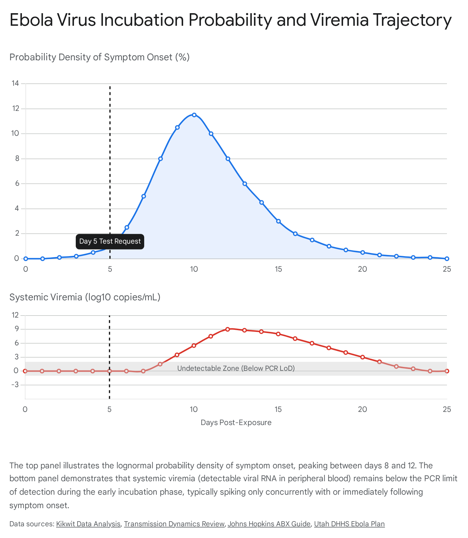
Epidemiological modeling of historical Ebola outbreaks, utilizing maximum likelihood inference and extensive contact-tracing datasets, indicates that the incubation period does not follow a normal distribution. Instead, the time from infection to illness onset is best fitted to a lognormal or Weibull distribution 2829.
Analyses of outbreaks involving the Zaire and Sudan species reveal specific statistical parameters for these distributions. For example, during the 1995 Kikwit outbreak of Zaire ebolavirus, the mean incubation period was estimated to be 12.7 days, with a standard deviation of 4.31 days 282930. Other models and distinct viral strains frequently cite an average incubation of 8 to 10 days, though the overarching shape of the probability curve remains heavily right-skewed 213132.
The cumulative distribution function of these epidemiological models reveals the following probabilistic landscape for symptom onset: * Days 1 to 4: An extremely low probability of symptom onset. The virus is undergoing primary replication in local tissues. * Day 5: The probability density begins to rise, but the vast majority of infected individuals have not yet developed any symptoms. * Days 8 to 12: The statistical mode and median of the distribution, representing the most common window for initial symptom presentation 212931. * Days 15 to 21: The long tail of the distribution, capturing late-onset cases. * Beyond Day 21: Mathematical density functions suggest that a small fraction of cases (estimated at approximately 4.1% in certain historical Zaire ebolavirus models) may exhibit an incubation period extending marginally beyond 21 days 2830. To reduce the risk of missed cases to below 1%, an incubation parameter of 25 days would theoretically be required, though 21 days remains the globally accepted maximum standard for practical quarantine, monitoring, and travel restriction protocols 182830.
If a traveler is evaluated by a clinician on day 5 post-exposure, they have only traversed the initial, low-probability tail of the incubation curve. The highest-risk window for symptom manifestation still lies 3 to 7 days ahead of them.
Clinical Progression and the Onset of Systemic Viremia
The progression of Ebola virus disease is deeply intertwined with the trajectory of systemic viremia. Understanding the clinical phases of the disease elucidates exactly when the virus begins shedding into the peripheral bloodstream in detectable quantities.
The Phases of Clinical Presentation
Once the incubation period concludes, the disease typically manifests in sequential phases, shifting from non-specific, flu-like presentations to severe gastrointestinal and hemorrhagic complications. Table 2 outlines this clinical progression and correlates it directly with the expected levels of systemic viremia.
| Phase of Infection | Timeframe (Post-Exposure) | Clinical Signs and Symptoms | Systemic Viremia Status |
|---|---|---|---|
| Incubation Phase | Days 2 to 21 (Average 8 - 10 days) 2131 | Asymptomatic. No clinical signs present. 21 | Undetectable in peripheral blood. Virus restricted to lymphatics/solid organs. 2233 |
| Early Prodromal ("Dry") Phase | Days 1 to 3 post-symptom onset | Abrupt onset of high fever (≥38.5°C), severe headache, myalgia, arthralgia, fatigue, sore throat, and chills. Easily misdiagnosed as malaria or influenza. 20313234 | Low to moderate. Rapid exponential rise begins. May still yield false-negative RT-PCR on day 1 or 2 of symptoms. 3335 |
| Mid-Late ("Wet") Phase | Days 4 to 7 post-symptom onset | Severe watery diarrhea, nausea, vomiting, abdominal pain, chest pain, maculopapular rash, conjunctival injection. 203132 | Very High. Peak viral loads frequently reaching $10^{8}$ to $10^{9}$ copies/mL of serum. 3637 |
| Late Severe Phase | > Day 7 post-symptom onset | Internal and external hemorrhage (petechiae, mucosal bleeding, bloody stool), multiorgan failure, hypovolemic shock, confusion, coma, and potential death. 203132 | Sustained High or Declining. High viral load at presentation correlates strongly with mortality. 3738 |
Viral Spillover into the Bloodstream
As indicated in Table 2, during the entirety of the incubation period - including day 5 post-exposure - Ebola virus RNA remains strictly below the limit of detection of sensitive molecular assays 33. The virus does not leak into the peripheral bloodstream in detectable quantities until the localized infection in the lymph nodes, liver, and spleen overwhelms local cellular defenses and begins driving massive cellular necrosis 2226.
When systemic spillover finally occurs, the viral load in the blood rises exponentially over a remarkably short time interval 33. Viral levels in symptomatic cases can rapidly escalate from undetectable to astronomical numbers within 48 to 72 hours 3637. Because this massive viral replication closely aligns with, or occasionally slightly trails, the onset of the early prodromal "dry" symptoms, blood drawn from an asymptomatic individual on day 5 will almost certainly test negative, failing to reflect the silent, accelerating replication occurring within their solid organs 32343839.
Analytical Sensitivity of Diagnostic Platforms
The asymptomatic traveler's request for a diagnostic test presumes that modern RT-PCR technology possesses the analytical sensitivity necessary to detect minute, pre-symptomatic traces of the virus in the blood. While modern assays are indeed highly sensitive, they are ultimately constrained by the biological absence of the target analyte in the sampled matrix.
RT-PCR Technologies and Limits of Detection
RT-PCR serves as the benchmark standard and primary diagnostic modality for Ebola virus disease due to its high gene- and species-specificity 394041. These molecular assays utilize specific primers and probes to target highly conserved regions of the filovirus genome, most commonly the nucleoprotein (NP) gene, the glycoprotein (GP) gene, and the matrix protein (VP40) gene 394042.
The Limit of Detection (LoD) represents the lowest concentration of viral RNA that an assay can reliably detect, typically calculated using Probit analysis to determine the concentration at which 95% of replicates test positive 36. Table 3 compares the analytical sensitivity of several prominent diagnostic platforms utilized in field and clinical settings.
| Diagnostic Platform | Target Gene(s) | Estimated Limit of Detection (LoD) | Processing Turnaround Time | Operational Characteristics |
|---|---|---|---|---|
| Cepheid Xpert® Ebola Assay | NP and GP genes | 73 copies/mL (inactivated) to 232 copies/mL (RNA) 36 | ~100 minutes 36 | Fully automated, single-cartridge point-of-care (POC) system; highly sensitive due to dual targets 3642. |
| Altona RealStar® Ebolavirus RT-PCR | L gene (Polymerase) | 509 copies/mL (95% CI: 411 - 722 copies/mL) 36 | Several hours | Conventional high-throughput laboratory assay requiring manual or automated separate RNA extraction 36. |
| BioFire FilmArray BioThreat-E | Multiplex (Unspecified EBOV targets) | Inconsistent detection below 4,850 copies/mL 36 | ~1 hour 43 | Multiplex panel identifying various biothreats; field-deployable but potentially less sensitive at trace viral loads 364344. |
| Co-Diagnostics BDBV Assay (2026 Update) | Bundibugyo specific | Technical data undergoing validation | Rapid POC | Newly developed decentralized PCR architecture targeting the specific 2026 Bundibugyo outbreak strain; currently under regulatory review 14950. |
Despite the extraordinary analytical sensitivity of systems like the GeneXpert assay - capable of detecting as few as 73 copies of viral RNA per milliliter of blood - clinical sensitivity is entirely dependent on the viral kinetics of the patient at the exact moment the venipuncture is performed 343642. The most technologically advanced assay will return a negative result if the virus has not yet entered the compartment being sampled.
The Target Gene Discrepancy
Extensive external evaluations of RT-PCR assays demonstrate that targeting the NP gene consistently yields higher analytical sensitivity than targeting the GP or VP40 genes 3940. Specifically, NP gene assays achieve detection limits as low as 1 to 10 RNA copies/μL for Zaire ebolavirus, whereas GP-gene specific assays frequently exhibit higher limits of detection ranging from $10^{2}$ to $10^{3}$ copies/μL 3940.
This discrepancy is a direct result of the virus's sequential transcription mechanism. During viral replication, mRNA transcription gradients inherently produce a greater number of copies of genes located closer to the 3' end of the genome. The NP gene is located closer to the 3' leader sequence, resulting in significantly higher transcript abundance compared to downstream genes 2726. Consequently, during the analysis of infectious particles, researchers have noted that while one copy of the NP gene is structurally included in each infectious virion, the overall abundance of NP mRNA in infected cells provides a robust target for early molecular detection 39. However, even with the deployment of optimal, highly sensitive NP-targeted assays, detection still fundamentally requires the virus or its transcripts to be physically present in the venous blood sample 3940.
The False-Negative Phenomenon During Incubation
The defining biological reason a day 5 RT-PCR test cannot clear an asymptomatic traveler is the temporal delay between cellular infection and systemic viremia, a phenomenon that guarantees a high rate of false-negative diagnostic results during the incubation period.
Mathematical Modeling of Diagnostic Sensitivity
A negative RT-PCR result during the incubation period is appropriately classified as a false negative relative to the patient's true systemic infection status 3335. Mathematical compartmental models, developed to track the transmission dynamics of Ebola virus outbreaks, divide populations into Susceptible, Exposed (incubating), and Infected (symptomatic/infectious) categories 35.
These rigorous epidemiological models indicate that the sensitivity of RT-PCR testing during the "Exposed" or incubation phase is effectively zero, conservatively estimated to range between 0% and 5% 35. Conversely, the RT-PCR sensitivity for infectious individuals tested 72 hours after symptom onset approaches 95% to 100% 35. Relying on a diagnostic test that carries a 95% to 100% false-negative rate during the specific timeframe in which it is administered provides absolutely zero diagnostic reassurance.
Clinical Evidence of Pre-Symptomatic Evasion
Clinical case reports meticulously document this diagnostic reality. In one notable instance, a pediatric patient subjected to quantitative RT-PCR testing during the late incubation phase returned negative results from blood drawn on day 4 post-exposure, only to test strongly positive 48 hours later as acute febrile symptoms progressed 33. This case reinforces the evidence that circulating virus remains firmly below the level of detection of sensitive molecular assays during incubation, and it highlights the shockingly rapid rate at which the virus eventually proliferates in the blood over a short time interval 33.
Consequently, diagnostic guidelines, including instructions for use authorized by the US Food and Drug Administration (FDA) for EBOV RT-PCR assays, explicitly warn clinicians that a negative test result during the early stages of disease cannot be relied upon to rule out infection 334546. When a diagnostic test returns negative, the possibility of a false negative must be heavily weighted against the patient's epidemiological exposure risk 4546.
Public Health Protocols for Contact Clearance
Given the total unreliability of molecular diagnostics during the incubation period, global public health frameworks uniformly reject early asymptomatic testing as a valid mechanism for viral clearance.
The Mandatory 21-Day Observation Standard
The protocols established and enforced by the World Health Organization, Africa CDC, and the US CDC dictate that any individual returning from an active outbreak zone, or possessing a known epidemiological risk factor, must undergo continuous monitoring for the full 21-day incubation period 7819.
For example, in response to the 2026 Bundibugyo outbreak in the Democratic Republic of the Congo and Uganda, the CDC utilized Title 42 regulatory authority to enforce enhanced public health screening, contact tracing coordination, and strict entry restrictions 818. During the required 21-day period following their departure from the affected region, the traveler is legally and medically obligated to monitor their physiological status, explicitly checking for initial "dry" symptoms (fever, chills, myalgia) and subsequent "wet" symptoms (diarrhea, vomiting, unexplained bleeding) 173247.
Standard operating procedures for contact clearance explicitly state that if a contact remains entirely afebrile and asymptomatic for 21 days following their last potential exposure, surveillance can be discontinued, and the individual is officially cleared 819. Clearance is granted strictly based on the passage of time without symptom onset, never on the basis of a negative laboratory test obtained during the incubation window.
Testing Protocols Upon Symptom Onset
Molecular testing is reserved strictly for individuals who develop clinical signs compatible with EVD 454748. Should the returning traveler develop a sudden high-grade fever on day 8, an RT-PCR test is immediately indicated 213247.
Yet, even when symptoms physically manifest, the kinetics of the virus dictate extreme diagnostic caution. The US CDC mandates that if a patient presenting with compatible symptoms and exposure history tests negative on a blood specimen collected less than 72 hours after symptom onset, the patient cannot be cleared and must remain isolated in a healthcare facility 1735. A second confirmatory RT-PCR test must be performed on an entirely new blood specimen collected $\geq$ 72 hours after symptom onset to definitively rule out the disease 1735.
The logical conclusion is stark: if the most sensitive diagnostic test available cannot definitively rule out the virus 48 hours after symptoms begin due to low initial viral load, it is a biological impossibility for that same test to rule it out 3 to 7 days before symptoms begin.
The Danger of Reassuring Asymptomatic Travelers
Performing an RT-PCR test on a highly anxious, asymptomatic traveler on day 5 is not only medically futile but represents a severe public health hazard. Providing a negative laboratory result to the traveler is highly likely to induce a false sense of security 2433. Believing they have been biologically "cleared" by a molecular test, the traveler may cease their daily temperature monitoring, ignore subsequent early symptoms, and break quarantine or travel restriction protocols 2438.
Individuals infected with Ebola virus become highly contagious to others only once symptoms begin to manifest, with infectivity rising dramatically as gastrointestinal symptoms and hemorrhage develop 242738. The false reassurance provided by an unwarranted day 5 PCR test directly heightens the risk of secondary community transmission by delaying isolation when true symptoms ultimately arise 243538.
Experimental Approaches to Early Detection
While current standard-of-care RT-PCR on whole blood or serum cannot clear an asymptomatic patient, ongoing advanced research aims to narrow the detection gap during the silent incubation period.
Peripheral Blood Mononuclear Cell (PBMC) Enrichment
One novel avenue of diagnostic research involves bypassing the cell-free serum to target the virus within the specific cellular compartments it utilizes for early replication. Because the Ebola virus initially targets macrophages and monocytes, isolating peripheral blood mononuclear cells (PBMCs) from whole blood via centrifugation may provide an enriched, highly concentrated source of viral proteins long before widespread systemic viremia occurs 34.
In experimental animal models utilizing liquid chromatography coupled with tandem mass spectrometry (LC-MS/MS), targeted viral proteins (specifically VP40 and the nucleoprotein) were successfully detected in PBMCs concurrently with the very first day of detectable viremia 34. This research suggests that high-sensitivity immunoassays targeting specifically enriched PBMC populations might eventually offer a slight temporal diagnostic advantage over standard serum RT-PCR 34. However, this methodology remains highly experimental, requires complex mass spectrometry infrastructure, and is not currently authorized or applicable for clinical clearance in human travelers.
Physiological Telemetry and Machine Learning
A non-biochemical approach to pre-symptomatic detection bypasses molecular biology entirely, relying instead on high-resolution physiological telemetry 49. Researchers have applied machine learning algorithms - specifically supervised random forest classification methodologies - to analyze subtle, sub-clinical physiological markers in non-human primate models exposed to filoviruses 49.
By continuously monitoring micro-fluctuations in heart rate, subtle variations in core temperature, and disruptions to normal diurnal rhythms, these algorithms have demonstrated the remarkable ability to detect the asymptomatic state of filovirus infection an average of 52 hours ($\pm$ 14 hours) before the onset of clinical fever 49. While successfully extending this telemetry capability to wearable, non-invasive sensors for humans could revolutionize contact monitoring and healthcare worker safety during active outbreaks, it currently serves as a conceptual early warning system designed to trigger isolation protocols, rather than a definitive diagnostic tool capable of clearing a patient 49.
Addressing Misconceptions in Returning Travelers
The request for a day 5 PCR test by a perfectly healthy traveler is often driven by a fundamental misunderstanding of viral transmission mechanics, exacerbated by intense anxiety. Clinical evaluations of such patients must prioritize risk communication and education to ensure compliance with the 21-day protocol.
Common misconceptions during outbreaks include the belief that Ebola virus is airborne, waterborne, or transmissible by asymptomatic carriers through casual contact 245051. In reality, Ebola virus requires a live host and is transmitted exclusively through direct physical contact with the bodily fluids of a person who is actively exhibiting symptoms 242751.
A person incubating the virus on day 5, who feels perfectly healthy, is entirely non-infectious to those around them 2751. Casual social contact, including sharing airspace or shaking hands with an asymptomatic individual, does not spread the virus 2751. Educating the traveler that their current asymptomatic status protects their family and community - and that continued monitoring is the only way to maintain that safety - is a vastly more effective clinical intervention than ordering a medically useless RT-PCR test 242751.
Conclusion
In direct response to the clinical query regarding the asymptomatic traveler returning from an active outbreak zone: If the traveler undergoes an Ebola virus RT-PCR test on day 5 post-exposure and the result returns negative, they are definitively not cleared of the virus.
The negative result on day 5 indicates only that systemic viremia has not yet reached the analytical limit of detection of the assay within the peripheral venous blood. The biological pathogenesis of the virus - specifically its sequestration and highly localized replication within the lymphatic tissues, solid organs, and intracellular viral factories during the 2 to 21-day incubation period - ensures that viral RNA remains undetectable in serum until immediately before, or shortly after, the onset of clinical symptoms 22232534. Rigorous mathematical modeling and clinical case studies confirm that RT-PCR testing during the incubation phase carries a sensitivity approaching zero 3335.
Administering diagnostic tests to asymptomatic individuals provides zero actionable diagnostic value and poses a severe public health risk by potentially inducing a false sense of security, which may lead to catastrophic non-compliance with critical symptom monitoring protocols 243338. In accordance with universal, standardized guidelines enforced by the World Health Organization, the Africa Centres for Disease Control and Prevention, and the US Centers for Disease Control and Prevention, the only mechanism by which a traveler can be definitively cleared of the virus is to complete the mandatory 21-day symptom monitoring period without developing any clinical signs of Ebola virus disease 81319. Should symptoms subsequently arise, sequential RT-PCR testing, strictly including blood samples drawn $\geq$ 72 hours post-symptom onset, is required to definitively diagnose or rule out the infection 1735.