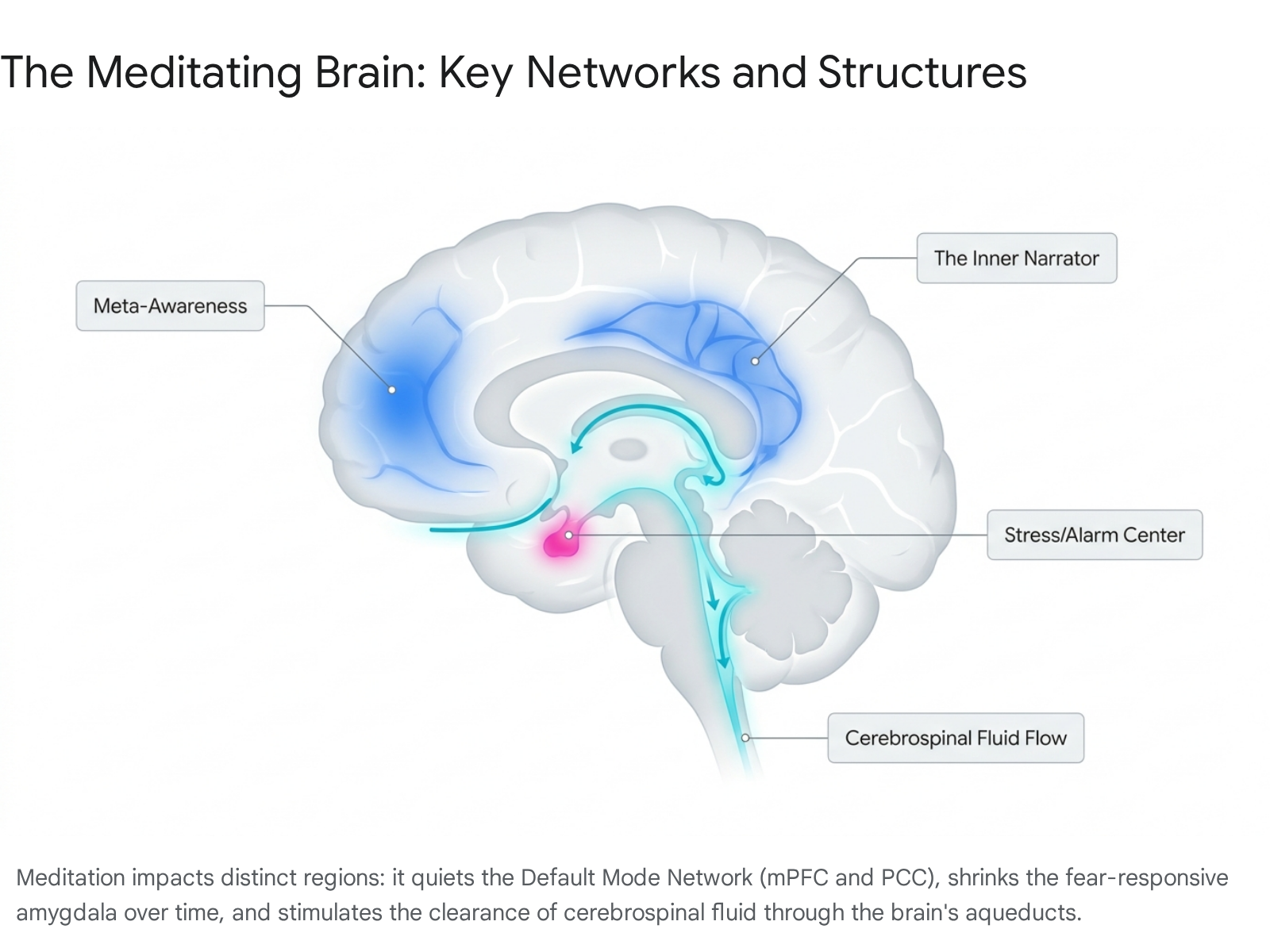
What Happens in Your Brain When You Meditate
Meditation immediately shifts the brain's electrical activity, quieting the neural networks responsible for mind-wandering and stress while promoting a state of relaxed, highly focused alertness. Over weeks and years of consistent practice, these functional changes lower systemic cortisol, alter the flow of cerebrospinal fluid to clear toxic metabolic waste, and can even preserve cortical volume against the natural aging process. However, recent rigorous neuroimaging has shown that the profound psychological benefits of beginner meditation are driven by functional rewiring - how neurons fire and communicate - rather than immediate, massive structural growth in brain tissue.
The First Ten Minutes: Real-Time Brainwave Shifts
A pervasive misconception about meditation is that it requires months of grueling isolation to produce any tangible neurobiological effect. In reality, the human brain begins to alter its operational frequencies almost the moment a practitioner closes their eyes and begins tracking their breath. The transition from an ordinary waking state to a meditative state is remarkably swift, measurable, and highly structured across different demographics.
Traditionally, contemplative neuroscience relied on comparing a broad baseline state of "rest" against an extended state of "meditation," failing to capture the moment-to-moment temporal dynamics of the brain as it drops into practice 1. That changed with a 2026 study published in the journal Mindfulness, which utilized 128-channel high-density electroencephalography (EEG) to map exactly when and how the brain changes during a ten-minute Isha Yoga breath-watching session 23. The researchers observed participants ranging from total novices to advanced practitioners. They utilized a mathematical approach known as threshold-free cluster enhancement to analyze data across all sensors simultaneously, preventing false positives and revealing the precise timeline of neurological shifts 13.
The data proved striking. Regardless of prior experience, significant, measurable changes in brainwave activity began to emerge within just two to three minutes of initiating the meditation 14. These alterations formed a distinct pattern that peaked in intensity between the seven- and ten-minute marks of the session 12.
To grasp the implications of these changes, one must understand the distinct frequencies at which neural populations oscillate. During these initial minutes of meditation, Alpha power (8 - 12 Hz) steadily and rapidly increased 4. Alpha waves serve as the brain's signature for wakeful relaxation - the mental state associated with quiet focus, free from acute anxiety or external distraction. Simultaneously, Theta power (4 - 8 Hz) rose, which is heavily associated with deep internalized attention, creativity, and the cognitive absorption that seasoned meditators describe as "going inward" 4.
Interestingly, these relaxing frequencies were paired with an increase in Beta1 power (13 - 20 Hz), a brainwave state that reflects alert, engaged focus. The simultaneous elevation of Alpha, Theta, and Beta1 produces a hybrid neurobiological state that researchers term "relaxed alertness," wherein the mind is deeply calm yet sharply awake 14. At the very same time, low-frequency Delta waves and high-frequency Gamma1 waves - which are heavily correlated with drowsiness, erratic mind-wandering, and low alertness - experienced a steady decrease 12. Within merely three minutes, the brain physically reorganized its electrical firing to become simultaneously more focused, more relaxed, and less prone to distraction 4.
While both beginners and experts experienced this timeline, the 2026 data revealed a crucial difference in how an expert brain rests. Advanced meditators demonstrated a highly distinct brainwave signature from the very first thirty seconds of the practice, exhibiting significantly elevated Theta and Theta-Alpha power compared to the other groups 34. This early, intense burst of internalized attention suggests that thousands of hours of lifetime practice fundamentally condition the brain's default resting state, allowing veterans to plunge into deep neurological shifts almost instantaneously rather than requiring a prolonged ramp-up period 14.
Deep Limbic Evidence in Absolute Novices
For decades, evaluating deep brain structures during meditation relied on functional Magnetic Resonance Imaging (fMRI), which measures blood oxygenation levels. However, placing a subject inside a loud, highly magnetic, claustrophobic fMRI tube often disrupts the very state of calm the researcher is attempting to measure 4. Scalp EEGs bypass the noise but struggle to capture electrical activity deep within the brain's limbic system, the evolutionary core responsible for raw emotional processing and memory.
In an effort to bypass these limitations, a groundbreaking 2025 study from the Icahn School of Medicine at Mount Sinai published in the Proceedings of the National Academy of Sciences (PNAS) utilized an unprecedented methodology. Researchers recruited neurosurgical patients with drug-resistant epilepsy who already had responsive neurostimulation systems surgically implanted directly into their brains 5. These chronic implants allowed the researchers to capture intracranial EEG recordings - direct, pinpoint electrical data from deep inside the amygdala and hippocampus - while the patients meditated.
The participants, all of whom were self-reported meditation novices, completed a simple five-minute audio-guided instruction 5. The intracranial recordings revealed that even during this brief, first-time practice, meditation induced immediate and profound changes in deep brain wave activity 5. The amygdala, often dubbed the brain's primal fear and alarm center, and the hippocampus, the critical hub for memory consolidation and emotional regulation, both altered their electrical firing patterns in real-time 5. This deep-tissue data confirms that the earliest moments of meditation do not merely induce superficial relaxation on the cortex; they immediately interface with the ancient, subcortical hardware responsible for emotional regulation and psychiatric resilience.
Silencing the Inner Narrator: The Default Mode Network
To fully appreciate why a ten-minute shift in Alpha and Theta waves produces such profound psychological relief, it is vital to examine what the human brain does when it is not meditating.
When an individual is awake but not engaged in a cognitively demanding, externally focused task, their brain does not simply power down. Instead, it defaults to a highly active, metabolically expensive web of interacting regions known as the Default Mode Network (DMN) 78. The primary anatomical hubs of the DMN include the medial prefrontal cortex (mPFC), the posterior cingulate cortex (PCC), the precuneus, and the parietal lobules 76.
Neuroscientists and contemplative scholars often characterize the DMN as the brain's "inner narrator" or "internal critic" 86. It is the network responsible for mind-wandering, self-referential thought, autobiographical memory recall, projecting anxieties into the future, and ruminating on the past 610. Under normal conditions, the default state of the human mind is to wander. Psychological research indicates that mind-wandering occupies roughly fifty percent of our waking hours and is robustly correlated with lower levels of subjective happiness and heightened baseline anxiety 7.
A hyperactive DMN acts as both the narrator and the editor of a person's mental autobiography. It can fuel cognitive biases, such as the overconfidence effect, while trapping individuals in cycles of half-formed certainties and self-referential stress 8. Modern psychiatry has closely linked the dysregulation and overactivation of the DMN to clinical conditions ranging from severe depression and anxiety to attention-deficit hyperactivity disorder (ADHD) and Alzheimer's Disease 7.
Meditation fundamentally disrupts this network. During focused attention practices - such as tracking the breath or repeating a mantra - the practitioner actively directs their cognitive resources away from self-referential narratives and toward a raw, sensory anchor. Functional neuroimaging reveals that as a meditator maintains this focus, the major nodes of the DMN experience relative deactivation 77. High-resolution 7-Tesla fMRI scans of beginner meditators performing focused attention tasks have demonstrated significant, widespread reductions in activity throughout the antero-medial prefrontal and posterior cingulate cortices 7.
Furthermore, as the DMN is quieted, other networks are upregulated. Brain states involving the dorsal attention regions occur more frequently, indicating improved attentional control even when the practitioner subsequently returns to a resting state 8. Connectivity analyses show stronger functional coupling between the posterior cingulate and the dorsolateral prefrontal cortex - regions implicated in cognitive control and conflict monitoring 7. Essentially, the act of meditating serves as resistance training for the mind. Every time a practitioner notices their attention wandering (an activation of the DMN) and intentionally redirects it to the breath, they strengthen the executive control networks capable of overriding the inner critic 713.
This neurological quieting produces a profound ripple effect on daily life. Practitioners spend less time locked in the frontal areas associated with analytical processing and rumination, and more time in networks synchronized with visual and sensory awareness 8. The result is a shift from abstract, anxiety-driven thinking toward embodied, present-moment sensory presence 8.
Mapping the Minimum Effective Dose
Understanding that the brain responds immediately to meditation naturally leads to a critical clinical question: How much daily practice is required to trigger lasting physiological and psychological adaptations?
Just as pharmacologists map the dosage requirements for medication, contemplative scientists have rigorously analyzed the "minimum effective dose" (MED) for mindfulness. The evidence reveals a clear dose-response relationship. Brief, consistent sessions are highly effective for protecting baseline cognitive function and buffering against daily stress. However, resolving severe clinical distress, reversing burnout, and driving deep shifts in generalized well-being require a significantly higher volume of practice.
The Dose-Response Matrix
When evaluating clinical trials, systematic reviews, and large-scale longitudinal studies, distinct tiers of neurological and psychological benefits align closely with specific practice durations.
| Daily Practice Dose | Primary Neurological & Psychological Outcomes | Key Scientific References & Findings |
|---|---|---|
| 10 to 13 Minutes | Reduces morning cortisol spikes by 15 - 25% over 8 weeks. Upgrades working memory, sharpens attention span, and begins to functionally deactivate the Default Mode Network. | Dr. Amishi Jha; 2019 Behavioural Brain Research. Splits into smaller 5-minute sessions remain effective. 1491610 |
| 20 to 30 Minutes | Triggers profound autonomic shifts. Modulates cerebrospinal fluid (CSF) dynamics for brain waste clearance. Produces moderate-effect improvements in clinical anxiety. | Vanderbilt PNAS 2025 CSF study. Standard minimum for establishing autonomic resilience. 111920 |
| 35 to 65 Minutes | Required for clinically meaningful, generalized shifts in overall well-being. Generates deep shifts in positive affect and begins to stabilize trait-level neuroplasticity. | Bowles & Van Dam 2025 prospective longitudinal study tracking 1,053 practitioners. 2112 |
| 50 to 80 Minutes | The threshold necessary to heavily attenuate severe clinical distress and drastically alter chronic mental health outcomes (depression, profound anxiety, burnout). | Bowles & Van Dam 2025. Aligns with intensive retreat protocols and clinical intervention upper bounds. 2123 |
Consistency Over Volume
For the vast majority of the population seeking cognitive enhancement and stress reduction, the most critical variable is not the length of the individual session, but the frequency of the habit.
In a landmark 2025 longitudinal study published in Applied Psychology: Health and Well-Being, researchers Bowles and Van Dam monitored 1,053 meditators across a diverse sample spanning North America, Europe, and Australia 1213. Tracking participants over a two-month prospective period with a two-to-four-year follow-up, they mapped the dose-response relationships between practice history, session length, and psychological outcomes.
The data decisively showed that while total accumulated practice time is important, practice frequency was a much stronger predictor of beneficial outcomes than session duration 1213. A practitioner who sits for ten minutes, six days a week, will likely derive far greater autonomic and attentional stability than one who sits for a single, sixty-minute session on a Sunday 910. Randomized controlled trials further corroborate this, revealing that distributing practice - such as splitting a twenty-minute requirement into two separate ten-minute blocks - yields identical reductions in psychological distress and anxiety as a single massed session 21.
Ultimately, the baseline for cognitive protection is low. A mere 12 minutes of focused attention training per day, maintained over several weeks, is sufficient to significantly upgrade working memory capacity, decrease self-reported mind-wandering, and measurably lower the body's circulating stress hormones 1421.
The Eight-Week Mark: Functional Rewiring and Autonomic Tone
If a practitioner maintains their minimum effective dose for approximately eight weeks - the standard duration of structured clinical protocols like Jon Kabat-Zinn's Mindfulness-Based Stress Reduction (MBSR) program - the temporary shifts in brainwaves and attention begin to hardwire into enduring physiological traits.
The Chemical and Autonomic Shift
The most rapidly detectable systemic changes occur within the body's endocrine and autonomic nervous systems. A 2024 meta-analysis encompassing 58 randomized controlled trials and over 3,500 participants confirmed that meditation significantly and reliably reduces circulating cortisol levels 1416. The effect size is particularly large regarding the "cortisol awakening response" - the surge of stress hormone that naturally occurs shortly after waking up 16. Eight weeks of daily mindfulness practice typically drives this morning cortisol spike down by 15 to 30 percent, translating practically to a massive reduction in morning anxiety and physiological dread 911.
In tandem with dropping cortisol, the body exhibits a pronounced increase in Heart Rate Variability (HRV) 1011. HRV is a vital biometric indicator of autonomic resilience, reflecting the balance between the sympathetic (fight-or-flight) and parasympathetic (rest-and-digest) nervous systems. Higher HRV denotes enhanced parasympathetic tone, meaning the brain and body possess a much greater capacity to recover swiftly from acute stressors and maintain a calm baseline 1011.
The Functional Connectivity Shift
Beneath the skull, eight weeks of practice functionally rewires how distinct brain regions communicate. Rather than acting as isolated hubs, the brain operates through sweeping networks. Neuroimaging demonstrates that mindfulness training enhances the microstructural integrity of white matter tracts, facilitating better communication between the prefrontal cortex - the seat of rational, executive control - and the parietal and limbic regions 1325.
As these communication pathways strengthen, the practitioner becomes highly efficient at processing sensory information while simultaneously downregulating emotional reactivity 25. For instance, MBSR training has been shown to increase functional connectivity between the right insula (associated with bodily awareness and interoception) and dorsolateral prefrontal regions 14. The brain learns to observe a stressor, process the physical sensation of anxiety in the body, and utilize executive control to inhibit a panicked reaction, resulting in what psychologists measure as enhanced emotional regulation and resilience 1115.
The Great Structural Brain Controversy
For the better part of the last decade, science media widely disseminated the claim that an eight-week mindfulness course physically grew the brain. Early, highly publicized pilot studies from prestigious institutions suggested that short-term MBSR interventions resulted in increased gray matter density in the hippocampus and measurable shrinkage of the amygdala.
However, as contemplative neuroscience matured, these initial claims faced intense methodological scrutiny. Early studies often suffered from small sample sizes, a lack of randomization, and critically, an absence of active control groups 161730.
In 2022, a watershed replication attempt led by Dr. Richard Davidson and Tammi Kral at the University of Wisconsin-Madison was published in Science Advances 3018. The researchers designed the largest and most rigorously controlled structural study to date. They pooled magnetic resonance imaging (MRI) data from 218 meditation-naive participants across two randomized controlled trials 1632. Subjects were assigned to either an 8-week MBSR program, a waitlist control, or - crucially - a highly matched active control known as the Health Education Program (HEP), which involved exercise, music therapy, and nutrition education 1617.
The results were definitive: the researchers found absolutely no evidence that eight weeks of MBSR produced neuroplastic changes in gray matter volume, gray matter density, or cortical thickness when compared to either control group 171832. The brain did not physically grow or shrink after two months of beginner meditation 1630.
The publication of these negative results forced a paradigm shift in the field. It highlighted that the undeniable, life-altering psychological benefits reported by MBSR graduates are overwhelmingly driven by functional changes (how the existing neural networks fire, synchronize, and communicate) and chemical changes (neurotransmitter and endocrine modulation), rather than immediate structural hypertrophy 173033.
Furthermore, the structural debate illuminated deeper methodological issues in the literature, including publication bias, where studies showing no effect were historically shelved, and instances of circular analysis 1719. Some recent meta-analyses attempting to salvage the short-term structural narrative have even faced retractions from major journals like Scientific Reports due to analytical flaws 151935. Ultimately, researchers now hypothesize that actually changing the physical macro-structure of the brain requires either an intervention significantly longer than eight weeks or a program singularly focused on one intense, specific form of practice rather than the broad, varied curriculum of standard MBSR 3020.
"Brain Washing": Cerebrospinal Fluid and Waste Clearance
While structural changes to gray matter require immense time, scientists have recently uncovered a profoundly rapid, physical change occurring in the brain's internal environment during meditation - one involving the fluids that surround and protect the organ.
Brain health relies heavily on the efficient circulation of cerebrospinal fluid (CSF) and interstitial fluid. These neurofluids circulate through regulated pathways in the central nervous system to clear out the toxic metabolic waste products that naturally accumulate as neurons fire during waking hours 2122. Efficient CSF flow is absolutely essential for flushing out problematic molecules, such as amyloid and tau proteins, the buildup of which is a primary driver of neurodegenerative conditions like Alzheimer's, Parkinson's, and Huntington's diseases 213940.
For decades, scientific consensus held that this deep "brain washing" process was strictly regulated by sleep 39. During deep, slow-wave sleep, the brain's fluid channels physically widen, allowing for a hyper-efficient washout of toxins via the glymphatic system. In contrast, aging and neurodegeneration are associated with sluggish, hyperdynamic, or "regurgitant" (backward-flowing) CSF circulation during waking life 22.
In late 2025, a groundbreaking study published in the Proceedings of the National Academy of Sciences (PNAS) by researchers at Vanderbilt University Medical Center challenged the assumption that this cleaning process was exclusive to sleep 2122. Led by Dr. Manus Donahue, the team hypothesized that because deep meditation shares several regulatory and arousal features with sleep, it might exert similar mechanical effects on brain fluids 2122.
Using advanced phase-contrast MRI and blood oxygenation level-dependent (BOLD) MRI, the researchers measured the absolute CSF flow and velocity through the cerebral aqueduct - a small, fluid-filled canal deep within the brain - in a cohort of highly adept meditators 2239. The participants were scanned while resting in a normal mind-wandering state, and then again while engaged in a focused-attention mindfulness practice tracking their breath 39.
The neuroimaging revealed that as participants slipped into focused-attention meditation, the motion of their CSF became significantly more efficient 21. Absolute CSF flow motion through the aqueduct decreased from an average of 4.60 mL/min down to 4.17 mL/min, driven largely by a steep reduction in harmful, regurgitant (backward-directed) fluid velocity 2223. At the same time, low-frequency CSF fluctuations increased, operating in an inverse rhythm to brain blood flow 2122.
These fluid dynamics tightly mirrored the restorative clearance patterns typically only observed during sleep, and operated in a manner directionally opposite to the fluid stagnation seen in aging brains 2122. The implications of the Vanderbilt study are vast: focused-attention meditation provides a rare, non-pharmacological waking state capable of actively modulating fluid dynamics 2223. Sitting in silent, self-guided breath awareness actively supports the brain's waste clearance systems, offering a profound physiological mechanism linking mental training to long-term neurological health and dementia prevention 204042.
Years of Practice: Anti-Aging and Brain Criticality
While eight weeks of practice may not grow gray matter, neuroimaging of long-term expert meditators - those with thousands of hours of practice spanning years or decades - paints an entirely different picture. Sustained, lifelong practice eventually does alter the physical morphology of the brain, offering a powerful neuroprotective shield against the ravages of aging.
Structural Preservation and Morphometry
A comprehensive anatomical likelihood estimation (ALE) meta-analysis of over twenty neuroimaging studies, examining roughly 300 experienced practitioners, confirmed that long-term meditation is consistently associated with altered brain structure across multiple key regions 24. These structural adaptations include:
- The Frontopolar Cortex (BA 10): Increased thickness in areas key to meta-awareness and higher-order executive function 24.
- The Insula and Sensory Cortices: Enhanced density in regions governing exteroceptive and interoceptive body awareness, tying into heightened empathy and emotional regulation 2425.
- The Hippocampus: Increased gray matter in this critical hub for memory consolidation, contextualized emotional learning, and spatial navigation. Because the hippocampus is uniquely vulnerable to the toxic effects of chronic stress hormones, this thickening acts as a deep protective buffer against trauma 1424.
- The Amygdala: While beginner courses may not shrink it, years of practice lead to reduced volume and reactivity in the amygdala, making the expert physically less susceptible to fear-based hijacking and autonomic panic 141645.
- The Superior Longitudinal Fasciculus and Corpus Callosum: Increased white matter integrity in these tracts facilitates superior intra- and interhemispheric communication across the brain 2425.
The cumulative effect of these structural changes is profound. In a 2025 randomized trial published in Scientific Reports, researchers utilized machine learning algorithms trained on gray matter volume, white matter, and glucose metabolism to estimate the "brain age" of older expert meditators (those with over 20 years of practice) against cognitively unimpaired older controls 2647. The models revealed that the long-term meditators exhibited significantly lower, "younger" brain ages 26. At chronological age 50, an expert meditator's brain is estimated to be, on average, 7.5 years younger than that of a matched non-meditator 25. This structural preservation was strongly mediated by both attentional mechanisms (higher volume in fronto-parietal areas) and positive psycho-affective factors 47.
Modulating Brain Criticality
Beyond mere volume, expert meditation alters the fundamental complex dynamics of how the brain operates as an entire system. A highly unique 2026 international study led by the Université de Montréal utilized magnetoencephalography (MEG) - a rare, non-invasive technology capable of tracking fine-grained brain dynamics with unparalleled temporal precision - to scan 12 monks from the Thai Forest Tradition 48. Together, these monks had accumulated over 15,000 hours of meditation each.
The researchers analyzed the monks' brains while they performed both focused attention (Samatha) and open-monitoring (Vipassana) meditation. The MEG scans revealed that meditation profoundly alters "brain criticality" 48. A concept borrowed from statistical physics, criticality refers to the optimal state of equilibrium a complex system maintains between rigid order and complete chaos 48.
The brain possesses a "sweet spot" of criticality. A brain with too much order lacks flexibility and adapts poorly to new stimuli, while a brain with too much chaos is prone to malfunction, as seen in epilepsy 48. The expert monks demonstrated a heightened, highly efficient state of criticality, modulating neural oscillations to achieve a massive increase in the complexity and adaptability of their brain activity 48. Contrary to the popular western notion that meditation is simply "thinking about nothing," the MEG data proves it is an intensely active, highly alert cerebral state that renders the brain vastly more flexible and efficient at processing reality 48.
Does the Type of Meditation Matter?
"Meditation" is an umbrella term encompassing dozens of historically and technically distinct cognitive exercises. Just as sprinting and weightlifting alter the body differently, different meditative practices engage unique neural networks and produce distinct neurobiological outcomes. A major meta-analysis of fMRI and neural imaging studies highlights the stark neurological differences between major traditions 25.
| Meditation Category | Common Examples | Primary Neural Mechanism & Psychological Effect |
|---|---|---|
| Focused Attention (FA) | Zen, early Vipassana, Samatha, Isha Yoga | Narrows the aperture of awareness. Strongly activates the prefrontal cortex, heavily deactivates the Default Mode Network, and increases high-frequency neural coherence. Excellent for building raw attentional capacity. 13254827 |
| Open Monitoring (OM) | Advanced Vipassana, Open Awareness | Broadens awareness to observe thoughts without attachment or judgment. Engages sensory cortices and the insula (interoception) while reducing cognitive control. Highly effective for emotional processing. 134850 |
| Compassion & Loving-Kindness | Metta, Tibetan Buddhist techniques | Cultivates altruism and empathy. Specifically activates the medial prefrontal cortex and emotional processing centers (amygdala, right temporoparietal junction) while decreasing implicit biases. 1325 |
| Non-Sleep Deep Rest (NSDR) | Yoga Nidra, Body Scans (secularized) | Strips spiritual context for deep autonomic relaxation. While traditional seated meditation rarely spikes dopamine, full NSDR protocols have been shown to uniquely increase dopamine reserves in the basal ganglia by up to 65%. 10 |
While secularized, modern interventions like Mindfulness-Based Stress Reduction (MBSR) often blend elements of FA and OM to treat clinical disorders, ancient practices like Vipassana originally aimed at fundamentally eradicating suffering through total presence of mind, leading to different long-term psychological profiles 51.
Methodological Limitations in Contemplative Neuroscience
While the data supporting meditation is increasingly robust, the field of contemplative neuroscience is still navigating significant methodological hurdles. A primary limitation is the nature of functional MRI testing itself. To capture high-resolution brain images, subjects are placed into a confined, highly magnetic tube while exposed to extremely loud, erratic mechanical noises 4.
Researchers have noted that this highly stressful, claustrophobic environment fundamentally interferes with a practitioner's ability to reach deep meditative states 4. Studies comparing simulated MRI environments to silent sitting found that practitioners in the scanner reported having much shallower meditation experiences, greater distraction, and higher skin conductance (a marker of physiological arousal) 4. While new ultra-high strength 7-Tesla fMRI machines improve signal quality, the environmental confounding factors remain a serious hurdle for capturing the true depth of advanced states like Samadhi or Jhana 752.
Furthermore, the field is reckoning with past frailties. Older studies were often plagued by small sample sizes, a lack of randomization, reliance on self-reporting, and publication bias, wherein studies showing no effect were quietly shelved 171928. Even cutting-edge attempts to close the loop between subjective experience and brain activity using real-time neurofeedback - such as feeding a meditator visual cues based on their posterior cingulate gamma activity - have shown mixed results, with researchers cautioning that muscle artifact contamination can easily skew high-frequency EEG readings 54. Consequently, while the broader trajectory of the science is incredibly promising, researchers continually caution against overhyping preliminary data into absolute medical certainties 195055.
Bottom line
From the very first minute of practice, meditation rapidly alters the brain's electrical frequencies, replacing erratic mind-wandering with a highly organized state of relaxed alertness that quiets the Default Mode Network. While popular claims that brief, eight-week beginner courses physically "grow" the brain are likely overstated, consistent daily practice creates undeniable functional and chemical shifts that lower stress hormones and even alter cerebrospinal fluid dynamics to wash metabolic waste from the brain. For those who sustain the practice over years, it acts as a profound neuroprotective shield, physically altering brain morphology to preserve memory, shrink fear centers, and keep the aging brain biologically younger and highly adaptable.