Neurological changes from sustained meditation practice
Foundations of Contemplative Neuroscience
The scientific investigation of meditation has transitioned from early electroencephalographic observations of ascetic monks to highly sophisticated, multi-modal neuroimaging studies exploring the physiological limits of human neuroplasticity. Contemplative neuroscience seeks to decode the measurable functional, structural, and neurochemical alterations that occur in the brain during active meditation, recognized as state effects, as well as the enduring changes resulting from sustained practice over time, recognized as trait effects 1. Originally rooted in ancient spiritual traditions such as Buddhism, Hinduism, and Taoism, meditation encompasses a heterogeneous family of mental training practices aimed at regulating attention, emotion, and self-awareness 23.
As clinical and psychological interest in mindfulness-based interventions has surged globally over the past two decades, so has the necessity to map these subjective states to objective neural correlates. Early research in the field predominantly sought to isolate the physiological benefits of relaxation; however, contemporary neuroimaging indicates that meditation alters large-scale brain network dynamics, modulates high-frequency brain oscillations, and fundamentally decouples sensory processing from affective reactivity 245. The application of functional magnetic resonance imaging (fMRI), magnetoencephalography (MEG), and intracranial electroencephalography (sEEG) has provided unprecedented spatial and temporal resolution into the meditating brain 67.
Despite these advancements, the field of contemplative neuroscience is currently undergoing a rigorous methodological reckoning. Early, highly publicized claims of profound structural brain changes resulting from short-term meditation are increasingly being challenged by highly controlled, large-sample randomized trials. This is prompting a critical shift in the scientific consensus toward understanding meditation primarily through the lens of dynamic functional connectivity and neurochemical modulation rather than static gray matter morphology 889.
Functional Neuroplasticity and Large-Scale Brain Networks
The most consistent neuroscientific findings regarding meditation relate to its immediate and long-term effects on the brain's functional architecture. Rather than operating as isolated anatomical regions, the brain functions through the continuous interaction of large-scale distributed networks. Meditation profoundly alters the interplay between the three core networks of the "triple network model": the Default Mode Network, the Central Executive Network, and the Salience Network 1011.
The Default Mode Network and Self-Referential Processing
The Default Mode Network (DMN), primarily comprising the medial prefrontal cortex (mPFC), the posterior cingulate cortex (PCC), the angular gyrus, and the medial temporal lobe, is highly active during periods of waking rest. It is the neurological substrate for mind-wandering, rumination, autobiographical memory retrieval, and self-referential thought 101214. Hyperactivation or hyperconnectivity within the DMN - particularly involving the subgenual anterior cingulate cortex (sgACC) - is a well-established neural correlate of major depressive disorder and anxiety, sustaining repetitive negative thought patterns 13.
A neurobiological hallmark of nearly all meditation practices, with the exception of automatic self-transcending techniques, is the suppression of DMN activity. During both focused attention and open monitoring meditation, fMRI reveals significant deactivation of the DMN compared to baseline resting states 214. By voluntarily redirecting attention away from spontaneous, self-referential narratives toward a designated anchor or open awareness, meditators actively inhibit the mPFC and PCC. In highly experienced practitioners, this suppression manifests as a trait effect, leading to a permanent reduction in baseline DMN activation. This chronic downregulation correlates behaviorally with decreased rumination, reduced clinical anxiety, and enhanced present-moment awareness 141516.
The Central Executive and Salience Networks
The Central Executive Network (CEN), anchored in the dorsolateral prefrontal cortex (dlPFC) and the posterior parietal cortex, is responsible for top-down cognitive control, sustained attention, working memory, and conflict resolution. The CEN and DMN typically operate antagonistically; when a cognitively demanding task activates the CEN, the DMN is concurrently suppressed 1017.
The Salience Network (SN), driven by the anterior insula and the dorsal anterior cingulate cortex (dACC), acts as the neurological switchboard between the CEN and DMN. It detects behaviorally relevant internal and external stimuli and subsequently recruits the CEN to focus on those stimuli while disengaging the DMN 1013. The accompanying schematic illustrates the anatomical nodes of these three networks and their functional interplay.
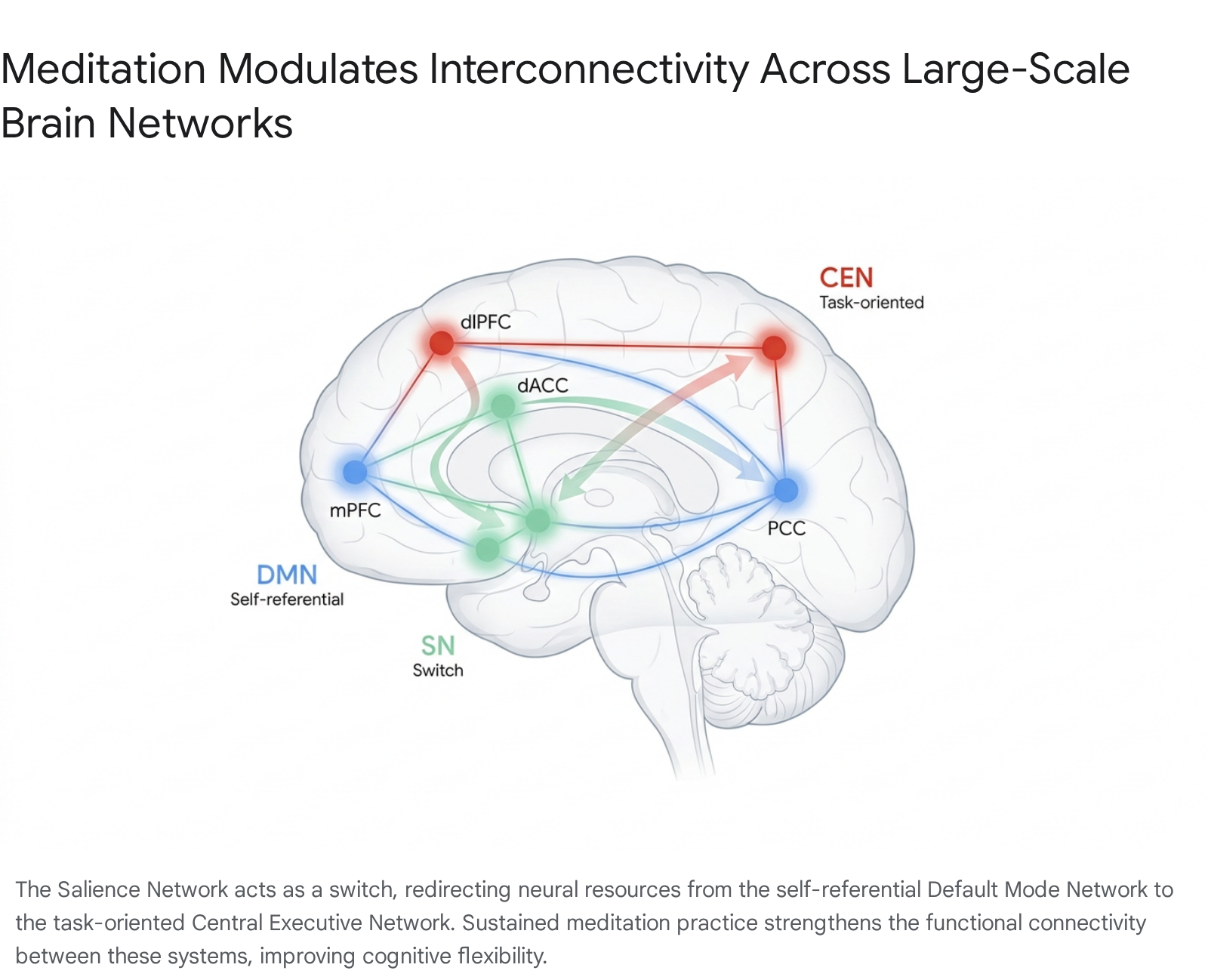
Neuroimaging demonstrates that extensive meditation training permanently enhances the functional connectivity between these macroscopic networks. Independent component analysis and sliding time window correlation analyses have shown that after as little as 31 days of mindfulness training, resting-state functional connectivity (rsFC) between the nodes of the SN and the DMN increases, alongside heightened connectivity between the SN and key regions of the CEN 11. This enhanced interconnectivity optimizes the brain's ability to seamlessly transition from internal introspection to task-oriented cognition, effectively strengthening the practitioner's metacognitive awareness and their capacity to rapidly disengage from affective distractions 1113.
Neural Signal Complexity and Temporal Dynamics
Beyond resting-state fMRI network analysis, advanced analytical techniques applied to magnetoencephalography (MEG) and electroencephalography (EEG) have revealed that meditation alters the temporal dynamics and mathematical complexity of neural oscillations. Neuroscientists measure brain signal complexity using metrics such as Lempel-Ziv complexity, spectral entropy, and long-range temporal correlations (LRTC) 718.
Research indicates that the meditative state is characterized by an overall increase in neural signal complexity compared to standard waking rest or mind-wandering, suggesting a richer, more diverse repertoire of neural micro-states 718. Simultaneously, expert meditators show attenuated long-range temporal correlations, particularly in the gamma band, indicating a breakdown of long-term predictive neural habits. This reduction in LRTCs aligns phenomenologically with the meditative goal of experiencing each moment anew, without the heavy imposition of prior cognitive schemas or predictive processing errors 718.
Comparative Neuroscience of Meditation Styles
Historically, scientific literature grouped diverse meditation techniques under the broad umbrella term "mindfulness," which occasionally led to conflicting or highly heterogeneous neurobiological data. Modern neuroscience recognizes that meditation is not a monolithic construct. Practices are now categorized into distinct phenomenological and neurological styles based on their cognitive mechanisms and resulting EEG signatures. The primary classifications include Focused Attention, Open Monitoring, Loving-Kindness, and Automatic Self-Transcending 1920.
| Meditation Category | Common Traditions | Primary Cognitive Mechanism | Functional Neural Correlates | Dominant EEG Signatures |
|---|---|---|---|---|
| Focused Attention Meditation (FAM) | Samatha, Zen, Vipassana (early stages) | Sustained concentration on a single object (e.g., breath); redirecting wandering attention. | Activation of dlPFC, ACC; suppression of DMN. Leftward frontal asymmetry. | High-frequency Beta-2 (20-30 Hz) and Gamma (30-50 Hz) 202122. |
| Open Monitoring Meditation (OMM) | Vipassana (advanced), Zazen, Mindfulness | Non-reactive, non-judgmental observation of moment-to-moment sensory and mental phenomena. | Deactivation of DMN; increased insula and somatosensory cortex activity. | Frontal Theta (5-8 Hz) and Occipital Gamma 2022. |
| Loving-Kindness & Compassion (LKM) | Metta, Tibetan Compassion | Cultivating altruistic intent and empathy for self and others through visualization and phrases. | Activation of superior parietal lobe, inferior frontal gyrus, and insular cortex 2324. | Variable, often localized Gamma synchrony in emotion-processing regions. |
| Automatic Self-Transcending (AST) | Transcendental Meditation (TM), Vedic Mantra | Effortless repetition of a mantra; transcends the cognitive control process itself. | DMN remains active; reduced amygdala reactivity 1420. | Alpha-1 (7-9 Hz), indicative of restful alertness and reduced mental effort 2021. |
Focused Attention Meditation
Focused Attention Meditation (FAM) serves as the foundational practice for many contemplative traditions, including Samatha and the early stages of Vipassana. It requires the top-down voluntary regulation of attention to maintain focus on a specific, chosen stimulus, most commonly the sensation of respiration 222526.
Neurobiologically, FAM heavily recruits the dorsal attention network and the anterior cingulate cortex (ACC). The ACC is critical for conflict monitoring and detecting when the mind has wandered away from the object of focus 2730. Functional imaging comparing expert Tibetan monks to meditation novices during FAM shows markedly different activation levels. Initially, novices show high ACC activation as they struggle to maintain focus. However, highly skilled monks exhibit efficient, sustained activation of the dlPFC and ACC with significantly less effort, highlighting the neuroplastic reinforcement of attentional control circuits 30. Electrophysiologically, FAM is characterized by enhanced Beta-2 (20-30 Hz) and Gamma (30-50 Hz) power, indicative of vigilant, active cognitive engagement 2021.
Open Monitoring Meditation
Open Monitoring Meditation (OMM), which includes advanced Vipassana and Zen (Zazen) practices, involves broadening the attentional spotlight to encompass all internal and external experiences without attachment, cognitive elaboration, or judgment 192526. Because OMM drops the requirement to rigidly focus on a single object, it reduces the demand on top-down executive control networks. Instead, it alters default mode activity by detaching the practitioner from autobiographical memory and reducing habitual emotional reactivity 31.
Studies utilizing the Stroop cognitive conflict task demonstrate that OMM practitioners exhibit highly efficient conflict resolution with less corresponding brain activation in prefrontal motor-control areas. This indicates that non-reactive monitoring and cognitive control become effortless, automatized traits in long-term practitioners 101219. Furthermore, EEG recordings of long-term Vipassana meditators reveal significantly increased occipital gamma power during meditation, a neural correlate associated with enhanced perceptual clarity and the rich binding of moment-to-moment sensory information 32.
Loving-Kindness and Compassion Meditation
Loving-Kindness Meditation (LKM) and related compassion practices diverge significantly from purely attentional training by explicitly engaging socio-emotional and empathy networks. The goal is to cognitively generate and sustain feelings of benevolence and empathy.
Long-term LKM practitioners display altered structure and function in the superior parietal lobe, the inferior frontal gyrus, the medial frontal lobe, and the insular cortex 24. These regions are integral to the brain's "theory of mind" network, affective empathy, and the translation of interoceptive visceral states into conscious emotional awareness. Unlike FAM and OMM, which primarily seek to dampen emotional reactivity or observe it neutrally, LKM actively upregulates specific emotional circuits, fostering structural neuroplasticity in areas dedicated to prosocial behavior and self-compassion 1624.
Automatic Self-Transcending and Mantra Meditation
Automatic Self-Transcending (AST) techniques, notably Transcendental Meditation (TM) and various Vedic mantra practices, utilize the silent, effortless repetition of a sound or phrase to guide the mind away from active, discursive thinking 3334.
Unlike FAM, which requires effortful concentration, or OMM, which requires active vigilant monitoring, AST is defined phenomenologically and neurologically by its lack of cognitive effort 20. Consequently, the DMN is not suppressed during TM practice; rather, it remains highly active while the brain enters a unique state of deep physiological relaxation. This state is characterized by globally coherent Alpha-1 waves (7-9 Hz), distinguishing it clearly from the high-frequency gamma activity of Buddhist practices or the delta waves of deep sleep 142034. Meta-analyses have shown that this specific neurophysiological profile is highly effective in reducing trait anxiety and fostering inner directedness 2021.
Structural Brain Alterations: Evidence and Controversies
While the functional and electrophysiological impacts of meditation are well documented and largely undisputed, the assertion that meditation physically alters the macro-structure of the brain remains one of the most heavily debated topics in contemporary neuroscience.
Early Findings on Gray Matter and Cortical Thickness
In the early 2000s, pioneering morphometric MRI studies suggested that sustained meditation practice induced robust, localized structural neuroplasticity. Seminal research found that experienced Insight (Vipassana) meditators possessed increased cortical thickness in the prefrontal cortex and the right anterior insula - regions critical for executive function, interoception, and sensory processing - compared to matched meditation-naïve controls 3228. Furthermore, these early studies reported that the differences in cortical thickness were most pronounced in older participants, suggesting that meditation might offset natural age-related cortical thinning and provide an "age-defying" neuroprotective effect 3229.
Subsequent meta-analyses of these early morphometric studies pointed to specific brain regions consistently showing increased gray matter density or volume in meditators. These included the hippocampus (vital for memory consolidation and context regulation), the orbitofrontal cortex (emotion regulation), the anterior cingulate cortex, and sensory cortices 142829. Conversely, regular meditation was widely reported to be associated with a reduction in the size and volume of the amygdala. This structural decrease correlated strongly with self-reported decreases in stress and fear responses, providing a neat neuroanatomical explanation for the psychological benefits of mindfulness 1530.
Methodological Limitations and the Replication Crisis
Despite the widespread popularization of these structural findings, rigorous methodological scrutiny has cast severe doubt on the premise that short-term meditation interventions can physically rewire brain macro-structure. The turning point in this neuroscientific discourse was a 2022 landmark study published in Science Advances by Kral, Davidson, and colleagues at the Center for Healthy Minds at the University of Wisconsin-Madison 89.
This study represented the largest and most rigorously controlled randomized controlled trial (RCT) on mindfulness and structural neuroplasticity to date. The researchers tested 218 meditation-naïve participants, assigning them to an 8-week Mindfulness-Based Stress Reduction (MBSR) course, a waitlist control group, or a validated, matched active control group (a non-mindfulness Health Enhancement Program) 931. Analyzing gray matter volume, gray matter density, and cortical thickness at both the whole-brain level and within specific pre-defined regions of interest (including the amygdala, hippocampus, and insula), the researchers found zero evidence of structural brain changes resulting from the MBSR program 89.
This prominent failure to replicate earlier claims highlighted severe methodological flaws that permeated the prior structural literature. Earlier positive findings were likely the result of small sample sizes, cross-sectional designs that could not rule out pre-existing brain differences in people drawn to meditation (self-selection bias), and reliance on circular statistical analysis 88928. Most critically, early studies often relied solely on waitlist controls. A waitlist control cannot account for the general psychological benefits of social engagement, expectation of relief, and active learning. When MBSR was compared to an active control program that matched the time, attention, and group dynamics of meditation training, the structural differences vanished 8931.
Recalibrating Structural Neuroplasticity Hypotheses
The scientific consensus is currently recalibrating in light of these highly powered replication failures. While lifelong, intensive meditation (e.g., thousands of hours accumulated over decades in monastic settings) may eventually yield macroscopic structural morphological changes, standard short-term interventions (such as 8-week MBSR programs or corporate mindfulness training) do not 816.
The behavioral and psychological benefits observed in short-term meditators are almost entirely driven by rapid changes in functional connectivity, dynamic network switching, and neurochemical balance (such as enhanced GABA and serotonin signaling), rather than gross structural neurogenesis or gray matter proliferation 83233. Evidence for structural change is now shifting away from gray matter volume toward investigations of white matter integrity, where diffusion tensor imaging (DTI) suggests that meditation may strengthen the axonal wiring and communication efficiency between different brain regions over extended periods of practice 434.
Clinical, Cognitive, and Physiological Implications
Even with the tempering of structural claims regarding short-term practice, the functional neural shifts induced by meditation drive significant, measurable clinical and psychological outcomes.
Emotion Regulation and the Limbic System
The primary clinical application for mindfulness involves the down-regulation of the hypothalamic-pituitary-adrenal (HPA) axis and the attenuation of limbic system reactivity 283033. While the physical macro-structure of the amygdala may not shrink in eight weeks, its functional activation profile alters rapidly and dramatically.
Groundbreaking intracranial electroencephalogram (sEEG) studies - which utilize electrodes implanted deep within the brain of epilepsy patients to bypass the spatial limitations of scalp EEG - have recorded immediate changes in wave activity within the amygdala and hippocampus during even a single, first-time audio-guided meditation session 6. By suppressing amygdala hyperactivity and strengthening top-down inhibitory control from the medial prefrontal cortex, meditation facilitates a shift toward parasympathetic nervous system dominance. This is reflected in clinical markers such as improved heart rate variability (HRV) and reduced salivary cortisol levels 15283334.
This altered limbic connectivity translates clinically into moderate reductions in anxiety, depression, and perceived stress. However, large-scale meta-analyses and systematic reviews consistently note that the efficacy of mindfulness for these psychiatric conditions is generally comparable to, rather than vastly superior to, other established active interventions like cognitive behavioral therapy or structured physical exercise 82835.
Interoception and Pain Modulation
One of the most robust and clinically valuable neurobiological findings is meditation's unique capacity to alter the perception of pain. When meditation-naïve individuals experience painful stimuli, the brain's sensory regions (primary and secondary somatosensory cortices) activate synchronously with affective regions (the anterior cingulate cortex, amygdala, and insula) 15. Consequently, pain is experienced simultaneously as a physical sensation and an aversive, highly distressing emotional threat.
In long-term meditators, functional neuroimaging reveals a profound functional decoupling of these discrete networks. During the application of thermal pain, expert practitioners show heightened, high-resolution activation in primary sensory processing areas, but drastically reduced activation in the affective appraisal networks (the amygdala, thalamus, and posterior cingulate cortex) 15.
Through somatic practices like Vipassana body-scanning, meditators train the right anterior insula to process interoceptive bodily signals objectively and with high precision. This neural decoupling enables practitioners to perceive the pure sensory intensity of a painful stimulus without generating the secondary cognitive and psychological suffering typically associated with it. In clinical and experimental settings, this functional reorganization has resulted in pain intensity and unpleasantness reductions of up to 40%, demonstrating unique mechanisms of analgesia distinct from placebo responses 228.
The Potential Adverse Effects of Meditation
While heavily promoted in mainstream culture as a universal panacea for mental health and cognitive enhancement, contemplative neuroscience and clinical psychology are increasingly documenting the potentially adverse psychological and neurological effects of meditation.
Because intensive meditation inherently deconstructs habitual self-referential processing, lowers psychological defense mechanisms, and dramatically alters baseline neural activity, it can precipitate acute distress in vulnerable individuals. Large-scale cross-sectional studies and systemic reviews indicate that a significant proportion of regular meditators experience unpleasant effects. One comprehensive study reported that over 60% of intensive retreat participants experienced at least one adverse psychological event 2835.
These adverse effects range from transient anxiety and heightened emotional sensitivity to severe panic, profound disorientation, and full-blown dissociation or psychosis 2835. Extended periods of Open Monitoring and deep introspection can inadvertently lead to the resurfacing of repressed childhood trauma or the destabilization of the ego structure, often without the protective clinical scaffolding provided by traditional psychotherapy 1535. Consequently, researchers strongly advocate for rigorous psychiatric screening for pre-existing vulnerabilities before individuals undertake intensive meditative interventions or multi-day silent retreats 152835.
Emerging Methodologies and Interdisciplinary Research
The next frontier of contemplative neuroscience involves integrating advanced biometrics, longitudinal tracking, and targeted neurotechnologies to better understand and optimize meditative states.
Neuroscience Research Initiatives in South Korea
Institutions in South Korea, such as the Korea Advanced Institute of Science and Technology (KAIST) and Seoul National University (SNU), are pioneering interdisciplinary approaches to meditation science. The KAIST Center for Contemplative Research integrates brain-computer interface technology, virtual reality, and neuroscience to study cognitive behavior 3637. SNU's Department of Psychology and Neuroscience Research Institute heavily utilize fMRI and genomic data to construct predictive computational models of the human mind under meditative states 3839.
Recent breakthroughs from these joint South Korean teams include the development of wireless, non-invasive near-infrared spectroscopy to monitor the glymphatic system - the brain's deep waste clearance mechanism - in real-time 40. Because meditation promotes deep parasympathetic relaxation, enhances slow-wave sleep architecture, and alters cerebral blood flow, researchers are actively investigating whether sustained meditative practice enhances the glymphatic clearance of amyloid-beta proteins during sleep. If confirmed, this functional physiological mechanism could position meditation as a vital preventative lifestyle intervention against neurodegenerative conditions like Alzheimer's disease 32840.
Mindfulness-Based Neurofeedback
Additionally, the commercialization and clinical exploration of mindfulness-based neurofeedback (mbNF) attempts to accelerate meditative proficiency by providing real-time auditory or visual feedback based on the user's specific brainwave states (primarily utilizing consumer-grade EEG devices) 4142. The theoretical objective is to help novices rapidly identify and sustain target states, such as elevated frontal theta or reduced DMN activation.
However, comprehensive systematic reviews and meta-analyses of mbNF randomized controlled trials reveal mixed outcomes. While these devices offer a modest reduction in general psychological distress, there is currently no robust evidence that they significantly improve objective cognitive function, physiological health, or absolute state mindfulness compared to active control conditions 4142. Furthermore, mechanistic modulation of the targeted neural mechanisms via commercial neurofeedback has not been conclusively proven in highly controlled within-participant designs, suggesting that traditional, unassisted meditation training remains the gold standard for inducing functional neuroplasticity 4142.
Conclusion
The neuroscience of meditation reveals a highly dynamic interplay between cognitive behavioral practice and large-scale brain network architecture. The measurable, verifiable effects of sustained meditation are best understood not as immediate structural neurogenesis or isolated volumetric growths, but as profoundly enhanced functional integration and dynamic network flexibility.
By modulating the interconnectivity of the Default Mode, Salience, and Central Executive networks, regulating high-frequency neural oscillations, and decoupling pure sensory processing from affective reactivity, meditation provides a robust empirical mechanism for emotional regulation, stress resilience, and clinical pain management. Moving forward, contemplative neuroscience is shedding its early reliance on underpowered structural claims in favor of highly rigorous, replicated functional data, paving the way for targeted, clinically validated, and neurologically precise contemplative therapeutics.