Microplastics in human organs and tissues
Introduction to the Human Microplastic Burden
The exponential acceleration of global plastic production over the past six decades has precipitated widespread environmental contamination. Historically, research into microplastics (MPs) and nanoplastics (NPs) - collectively referred to as MNPs - focused heavily on marine ecosystems, soil degradation, and atmospheric transport. However, research conducted between 2024 and 2026 represents a critical paradigm shift, transitioning the focus from environmental ecology to human physiology and internal bioaccumulation 1234. Advanced spectroscopic and mass-based analytical methods have recently established that these anthropogenic synthetic polymers do not merely pass through the human gastrointestinal and respiratory tracts. Instead, they actively translocate into systemic circulation and lodge deep within highly protected organs 5678.
Microplastics are generally defined as plastic fragments, fibers, and spheres measuring less than 5 millimeters (mm) 12. Nanoplastics represent a far more invasive sub-category, typically defined as particles smaller than 1 micrometer (μm) or 1,000 nanometers (nm), with some toxicological definitions restricting the upper limit to 100 nm 2. The distinction between microplastics and nanoplastics is not merely semantic; the size differential dictates fundamentally distinct physical and biological consequences. Due to their minute volume and high surface-area-to-volume ratio, nanoplastics are capable of penetrating biological membranes, circumventing cellular defenses, and exerting toxicological activity that is orders of magnitude higher than their larger microplastic counterparts 25910.
Humans are chronically exposed to these particles via three primary portals: inhalation of airborne particulate matter, ingestion of contaminated water and food, and dermal absorption 8111213. Airborne exposure includes synthetic textiles, indoor dust, and tire wear particles, while ingestion pathways are dominated by seafood, agricultural products grown in contaminated soil, and ultra-processed foods 81214. Current epidemiological and post-mortem studies aim to quantify this continuous exposure, map the specific organs where polymers accumulate, and determine the cellular damage and clinical health outcomes triggered by their persistent presence 1617.
Mechanisms of Exposure and Systemic Entry
Before accumulating in secondary organs, MNPs must navigate and breach the body's primary epithelial barriers. The efficiency of this translocation is heavily dependent on the physicochemical properties of the particles, particularly their size, shape, surface charge, and hydrophobicity 816.
Gastrointestinal and Pulmonary Absorption
Ingestion is widely considered the most significant route of human MNP exposure. While larger microplastics (>10 μm) generally fail to cross the intestinal epithelium and are excreted in feces, sub-micron particles exhibit substantial cellular uptake 78. The oral bioavailability of a 50 nm polystyrene nanoparticle is estimated to be ten to one hundred times greater than that of a multi-micrometer particle 28. In the gastrointestinal tract, MNPs are internalized predominantly via transcellular endocytosis or paracellular transport through the tight junctions of the intestinal epithelium 15.
Inhalation represents the second major exposure pathway. Airborne microplastics, generated from the friction of synthetic vehicle tires on road surfaces (TRWPs) and the weathering of textile fibers, are ubiquitous in both urban and indoor environments 1416. Research has identified that 97% of the plastic particles deposited in the human lungs range between 1 and 10 μm 9. Once inhaled, particles undergo phagocytosis by alveolar macrophages or cross the pulmonary epithelial barrier into the capillary network, directly entering systemic circulation 817.
Dermal Penetration
Dermal interaction with MNPs occurs primarily through contact with contaminated water, cosmetics, and personal care products 18. While the intact stratum corneum generally blocks particles larger than 100 nm, smaller nanoplastics can penetrate the epidermis and dermis 18. Experimental models utilizing human and murine skin have demonstrated that particles smaller than 1 μm, particularly 20 nm polystyrene nanoplastics, can concentrate in hair follicles and breach the dermal layers, although dermal uptake remains quantitatively lower than ingestion and inhalation 818.
Bioaccumulation in the Central Nervous System
The central nervous system, shielded by the highly selective blood-brain barrier (BBB), was traditionally presumed to be somewhat isolated from particulate contamination. This presumption was overturned by a landmark study published in Nature Medicine in 2025, which analyzed decedent brain, liver, and kidney tissues from autopsies conducted in 2016 and 2024 192021.
Disproportionate Organ Affinity and Temporal Growth
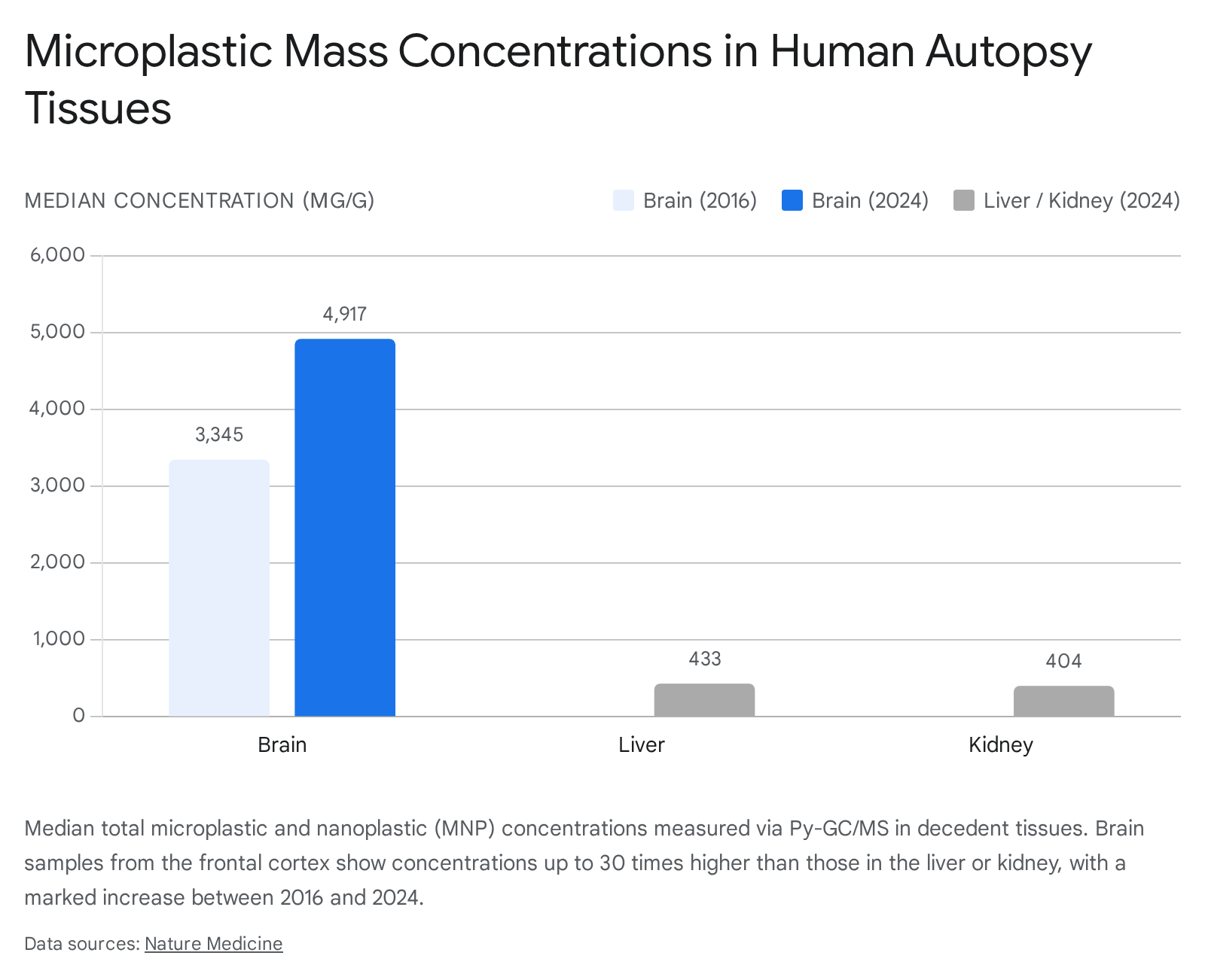
Researchers utilized pyrolysis gas chromatography-mass spectrometry (Py-GC/MS) to quantify the mass of plastic polymers per gram of human tissue. The findings demonstrated a significant, disproportionate accumulation of MNPs in the frontal cortex of the brain compared to peripheral filtration organs. In the 2024 sample cohort, brain tissue exhibited median plastic concentrations of 4,917 micrograms per gram (μg/g) 202122. This represented concentrations 7 to 30 times higher than the median concentrations observed in the liver (433 μg/g) and kidneys (404 μg/g) from the same individuals 2122.
Furthermore, a temporal comparison revealed an approximate 50% increase in brain MNP concentrations over an eight-year period, rising from a median of 3,345 μg/g in 2016 to 4,917 μg/g in 2024 20212223.
This upward trajectory closely mirrors the exponential increase in global plastic production and environmental fragmentation over the same timeframe. The researchers found no correlation between plastic concentrations and the age, sex, or race of the decedents, suggesting that internal accumulation tracks the intensity of recent environmental exposure rather than a slow, lifelong stockpiling process 202425.
Polymer Composition and the Dementia Correlation
The composition of the plastics found in the brain was highly specific. While liver and kidney tissues contained a wider array of polymers, the brain predominantly accumulated polyethylene (PE), which accounted for approximately 75% of the detected plastics 202123. Electron microscopy verified that these brain-bound polymers primarily existed as nanoscale shard-like fragments 202124.
The most provocative observation of the 2025 cohort involved donors with documented dementia diagnoses. Brains from dementia patients exhibited massive MNP concentrations - averaging 26,076 μg/g, which is significantly higher than age-matched healthy brains 2226. However, researchers enforce calibrated uncertainty regarding this metric: the data is strictly associative and does not establish that microplastics cause dementia 2123. Atrophy of brain tissue, impaired BBB integrity, and failing glymphatic clearance mechanisms are hallmarks of neurodegeneration; these pre-existing vulnerabilities likely make the dementia-afflicted brain highly porous, leading to secondary microplastic accumulation 2630.
Scientific Skepticism and Methodological Debate
It must be noted that these high-profile findings have encountered skepticism within the scientific community. Several chemists and environmental toxicologists have challenged the brain bioaccumulation studies, suggesting that the detections may be subject to false positives 3. Critics argue that certain fatty cells and lipid structures inherent to human brain tissue can produce spectral measurements that are easily confused with the signals emitted by common synthetic plastics 3. The inevitable use of post-mortem samples also prevents functional assessments of toxicity in living brains 27. Consequently, there is an international consensus that standardizing analytical methods and establishing forensic-level quality controls are prerequisites for validating these extreme tissue burdens 32829.
Translocation Mechanisms Across the Blood-Brain Barrier
The physiological mechanisms by which nanoplastics breach the blood-brain barrier remain a subject of intense investigation. The BBB is designed to block pathogens and chemicals, yet nanoplastics successfully infiltrate it through both structural disruption and molecular hijacking 1525.
Thermodynamic and Passive Permeation
Recent long-time-scale (over 28 microseconds) all-atom explicit solvent steered molecular dynamics have modeled the free energy required for the passive permeation of various polymers through the BBB 30. The data reveals that Polyethylene (PE), Polypropylene (PP), and Polystyrene (PS) exhibit a remarkable thermodynamic preference for entering the lipid bilayer of the BBB 30. This preference is attributed to their high hydrophobicity. These polymers can physically dissolve within the lipid structures of the barrier and subsequently exit into the brain parenchyma as dispersed polymer chains 30. Conversely, Polyethylene terephthalate (PET) was found to be energetically unfavored for passive BBB insertion 30.
Active Transport and Lipid Hijacking
Beyond passive permeation, biological mechanisms of BBB crossing include: 1. Disruption of Intercellular Junctions: MNPs can induce localized oxidative stress that degrades the tight junctions and adherens junctions between endothelial cells, artificially increasing barrier permeability and allowing paracellular transport 15. 2. Transcellular Endocytosis: Endothelial cells mistake the highly miniaturized nanoplastics for extracellular vesicles or nutrients, engulfing them via pinocytosis or clathrin-mediated endocytosis and transporting them across the cellular body 11215. 3. Translocation via Immune Cells: Often referred to as the "Trojan Horse" mechanism, MNPs are phagocytosed by peripheral macrophages. These immune cells subsequently migrate across the BBB, carrying the indigestible plastic burden directly into the central nervous system 131. 4. Lipid Hijacking: Researchers postulate that highly lipophilic nanoplastics bind to dietary fats and circulating lipids in the bloodstream. Because lipid transport pathways across the BBB are highly efficient, the attached plastics effectively hitch a ride into the brain tissue 2530.
The Biomolecular Protein Corona
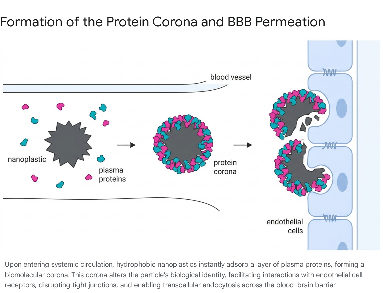
The physical and chemical behavior of a nanoplastic particle generated in a laboratory differs vastly from its behavior in a biological organism. Upon entering a biological fluid (such as blood plasma, interstitial fluid, or cellular cytoplasm), the pristine surface of a nanoplastic particle immediately attracts and adsorbs a complex layer of surrounding biomolecules - predominantly proteins, lipids, and metabolites 3233. This acquired coating is known as the "biomolecular corona" or "protein corona."
Altering Biological Identity and Toxicity
The protein corona functionally masks the underlying synthetic polymer, fundamentally altering its biological identity and governing its subsequent physiological fate 173233.
The composition of the corona is dictated by the particle's size, shape, surface charge, and hydrophobicity, as well as the specific biofluid it encounters 3234.
Highly hydrophobic nanoparticles strongly bind plasma proteins, which often leads to the unfolding and denaturation of the proteins, exposing hidden hydrophobic domains 32. This structural perturbation alters cellular recognition, signaling pathways, and immune responses 32. Positively charged nanoplastics preferentially attract negatively charged plasma proteins like serum albumin and fibrinogen 32.
Interestingly, while the corona facilitates cellular interaction, it can occasionally reduce immediate acute toxicity by acting as a buffer between the sharp synthetic surface and the cell membrane 33. In experimental studies evaluating BBB permeation, the presence of a robust biological corona on PS and PVC nanoplastics was shown to significantly reduce the raw quantity of particles that successfully entered the brain compared to pristine, uncoated particles 35.
Insights from Ecological Models
The importance of the corona is heavily supported by ecological toxicological assessments. Studies utilizing Daphnia magna (water fleas) exposed to nanoplastics coated with various biomolecules (fetal bovine serum, algae biomolecules, and Daphnia secretions) revealed that the corona composition directly influences toxicity and internalization 3642. Neonates exposed to bare, uncoated nanoplastics exhibited the highest mortality rates and the most severe swimming behavioral alterations, whereas those exposed to corona-coated plastics showed lower mortality but altered internalization pathways 36. Furthermore, exposure to the plastics triggered the organisms to secrete specific proteins aimed at mitigating oxidative stress, modifying the "eco-corona" in real-time 3642.
Pathological Impacts on the Cardiovascular and Reproductive Systems
The systemic accumulation of MNPs is associated with severe localized and cascading pathological effects, primarily driven by physical tissue irritation, chemical leaching, and oxidative stress.
Cardiovascular Disease and Prothrombotic Cascades
The link between microplastics and cardiovascular mortality gained unprecedented empirical support from the APAChE (Pollutants in the Atherosclerotic Plaque and Cardiovascular Events) study, published in the New England Journal of Medicine in 2024 373839. The prospective, multicenter observational study examined atherosclerotic plaque excised from the carotid arteries of 304 patients undergoing carotid endarterectomy 39.
Researchers detected measurable quantities of PE in the plaques of 58.4% of patients (mean level of 21.7 μg/mg of plaque), alongside PVC in 12.1% of patients 3739. The clinical outcomes were staggering: patients harboring detectable MNPs within their arterial atheroma faced a 4.53 hazard ratio - a 450% higher risk - of experiencing a composite endpoint event (myocardial infarction, stroke, or death from any cause) over a 34-month follow-up period compared to patients with plastic-free plaques 373839. In vivo data corroborates these findings, demonstrating that circulating MNPs induce endothelial dysfunction, disrupt cell membranes, and activate inflammatory cascades that create a highly prothrombotic state 263840.
Reproductive and Intergenerational Bioaccumulation
Reproductive toxicity represents another critical exposure vector. MNPs have been definitively located in human testicles, semen, amniotic fluid, and placentas, highlighting a threat to both fertility and fetal development 61947.
A 2026 study conducted in Indonesia identified nanoplastic contamination in human sperm and the amniotic fluid of pregnant women 47. From sperm samples, analysts recovered polyethylene particles ranging in size from 1.5 to 7.9 μm 47. Within the male reproductive tract, the presence of these nanoplastics is hypothesized to trigger oxidative stress that disrupts reproductive cell development, leading to diminished sperm quality, reduced motility, and abnormal morphology 1647.
In placentas, 2025 research indicated that MNPs were nearly universally present across tested samples 1619. Polyethylene constituted the majority of the particles (54%), followed by PVC 16. Crucially, elevated concentrations of placental microplastics correlated positively with preterm births, suggesting that the particles trigger localized inflammation and immune responses that destabilize the maternal-fetal barrier and induce premature labor 616.
| Tissue / Matrix | Major Polymers Detected | Particle Size Range | Observed or Hypothesized Health Implications |
|---|---|---|---|
| Brain (Frontal Cortex) | PE, PP, PVC | <0.02 μm to nanoscale shards | Deposition in cerebrovascular walls; associated with neuroinflammation and neurodegeneration 2024. |
| Carotid Plaque | PE, PVC | Bulk mass detected | 4.53x higher risk of myocardial infarction, stroke, or death over 34 months 39. |
| Blood | PE, Polyester, PET | 200 nm - 800 nm | Systemic transport vector; immune cell (macrophage) interaction 3147. |
| Placenta & Amniotic Fluid | PE, PP, PS, PVC | 2 μm - 300 μm | Disruption of maternal-fetal barrier, triggering inflammation linked to premature birth 61641. |
| Sperm / Testes | PE, PVC, Polystyrene | 1.5 μm - 7.9 μm | Reduced sperm motility, morphological abnormalities, oxidative destruction of germ cells 1647. |
Analytical Methodologies and Forensic Detection Frameworks
The identification of sub-micron plastic particles within lipid- and protein-rich human tissues pushes the absolute limits of modern analytical chemistry. In 2026, an international coalition of over 30 scientists explicitly called for the adoption of a "forensic science approach" to microplastic research to combat the high risk of false positives, matrix interferences, and cross-study inconsistencies 282942.
Human biofluids and tissues represent highly complex matrices. The inherent organic matter - fats, proteins, and cellular debris - often produces broad spectral features and background fluorescence that heavily overlap with the spectral bands of synthetic polymers 4344. Furthermore, biological samples are highly susceptible to airborne laboratory contamination, microfibers from technician clothing, and plastic degradation from standard laboratory storage equipment 2829. The forensic framework demands rigorous blank controls, optimized laser wavelength selections, and the mandatory use of at least two independent detection techniques (relying on different measurement principles) to definitively confirm the presence of a plastic polymer before reporting it as a positive finding 2942.
Categorization of Detection Technologies
Current detection methods are broadly categorized into mass-based approaches (which quantify total polymer weight) and particle-resolved approaches (which identify particle shape, size, and count) 44.
- Pyrolysis-Gas Chromatography/Mass Spectrometry (Py-GC/MS): A mass-based strategy that thermally degrades the tissue and plastic sample, analyzing the resulting gas fragments. It is highly sensitive and provides precise mass burdens (e.g., μg of PE per gram of tissue), which is ideal for exposure metrics and clinical correlations 4452. However, it destroys the sample entirely, preventing secondary morphological analysis, and can be susceptible to matrix interference if not calibrated strictly 1628.
- Fourier Transform Infrared Spectroscopy (μ-FTIR) and μ-Raman Microscopy: Particle-resolved vibrational techniques that rely on laser scattering to identify specific polymer bonds. While FTIR is generally limited to detecting particles larger than 10 μm, Raman can detect particles down to 1 μm 2544.
- Surface-Enhanced Raman Spectroscopy (SERS): SERS uses plasmonic substrates (like gold or silver nanoparticles) to enhance inherently weak Raman signals, allowing for the detection of nanoplastics down to 50 - 100 nm 245. While promising for nanoscale analytes, SERS is severely hampered in biological samples because the protein corona and surrounding organic matter interfere with the substrate, distorting peak positions and requiring complex multivariate deconvolution to isolate the true polymer spectrum 23443.
| Analytical Technique | Output Type | Approximate Size Limit | Primary Advantages | Limitations / Forensic Challenges |
|---|---|---|---|---|
| Py-GC/MS | Mass-based (μg/g) | Nanoscale (bulk mass) | High precision for total polymer weight; identifies specific chemical additives. | Destructive to the sample; cannot determine particle shape or exact size distribution 2844. |
| μ-FTIR | Particle-resolved | ~10 μm | Excellent for large microplastics and fibers; non-destructive. | Resolution limit excludes most nanoplastics; hindered by water interference in wet tissues 25. |
| μ-Raman | Particle-resolved | ~1 μm | Better spatial resolution than FTIR; effective for environmental samples. | Weak inherent scattering signals; highly susceptible to background fluorescence from biological tissues 24344. |
| SERS | Particle-resolved | ~50 nm | Amplifies Raman signals; allows identification of individual nanoplastics. | Complex sample preparation; severe signal interference from the protein corona and biomolecules 23443. |
Federal Funding, Policy, and Behavioral Interventions
Recognizing the escalating public health implications and the urgent need for standardized methodologies, the U.S. government shifted regulatory and funding frameworks significantly in early 2026.
Expanding Regulatory Scope and STOMP
The Environmental Protection Agency (EPA) took a historic step by adding microplastics to its Contaminant Candidate List (CCL 6) for the first time, establishing the necessary administrative groundwork for future drinking water regulations and mandatory municipal monitoring 465547.
Concurrently, the Department of Health and Human Services (HHS), through the Advanced Research Projects Agency for Health (ARPA-H), launched a $144 million federal research program known as STOMP (Systemic Targeting of MicroPlastics) 464858. The STOMP initiative represents a transition from purely observational research to active clinical intervention. It operates on a three-pronged approach - measure, target, and remove - with the explicit goals of developing a rapid, low-cost clinical diagnostic test to quantify individual microplastic body burdens, and subsequently exploring medical and biological therapies capable of safely eliminating these synthetic particles from the human body 46474858.
Efficacy of Behavioral and Dietary Interventions
While systemic solutions require protracted regulatory action and sweeping changes to global plastic production, recent clinical data indicates that individual behavioral interventions can successfully and rapidly reduce the body's internal plastic chemical burden.
The 2026 Plastic Exposure Reduction Transforms Health (PERTH) Trial, conducted by the University of Western Australia, provided the first robust quantitative data on behavioral mitigation 49. The randomized controlled trial monitored 211 healthy adults. The intervention group strictly adhered to a diet of food that had not contacted plastic during production or packaging, utilizing exclusively plastic-free kitchenware (such as stainless steel and wood), and substituting standard personal care products for low-plastic, fragrance-free alternatives 49.
The clinical results were immediate and statistically significant. Within just seven days of intervention, participants experienced a 44% to 60% reduction in urinary phthalates and a greater than 50% reduction in bisphenols (BPA/BPS) compared to the control group 49.
| Intervention Strategy | Mitigated Exposure Vector | Demonstrated Quantitative Impact |
|---|---|---|
| Avoiding Plastic Food Packaging | Chemical leaching into fresh produce and meats | Contributed to >50% drop in bisphenols within 7 days 49. |
| Eliminating Canned Foods/Beverages | BPA-coated resin liners inside aluminum cans | Reduced urinary endocrine disruptors . |
| Reducing Ultra-Processed Foods | Industrial manufacturing friction and multi-stage packaging | Decreased total plastic-associated chemical load . |
| Fragrance-Free Personal Care Items | Phthalates used in synthetic fragrances and plastic bottles | Mono-n-butyl phthalate dropped by 35% in 7 days . |
Conclusion
The period between 2024 and 2026 marked a critical evolutionary stage in environmental toxicology, confirming that microplastics and nanoplastics are ubiquitous not only in the external environment but deep within the human physiological architecture. The definitive quantification of mass plastic burdens in the frontal cortex of the brain, coupled with stark hazard ratios correlating arterial nanoplastics with cardiovascular mortality, underscores the severity of this chronic exposure.
While analytical limits persist - particularly regarding the spectral interference of the protein corona at the nanoscale - the adoption of strict forensic detection methodologies is actively stabilizing the data, separating genuine bioaccumulation from laboratory contamination. Regulatory agencies are beginning to pivot toward clinical diagnostics and targeted removal strategies, as evidenced by the $144 million STOMP initiative. However, until systemic global plastic reduction is achieved at the manufacturing level, robust behavioral interventions and targeted dietary modifications remain the most empirically validated mechanisms for minimizing individual toxicological risk.