Adult Neurogenesis in the Human Brain
For over a century, the foundational doctrine of neurobiology posited that the adult mammalian brain was a strictly post-mitotic organ, structurally immutable and incapable of generating new neurons after the conclusion of early developmental periods. This view, famously articulated by Santiago Ramón y Cajal, maintained that the neural pathways established during embryonic and early postnatal development were permanently fixed, with aging characterized solely by continuous neuronal attrition. However, pioneering autoradiographic studies by Joseph Altman in the 1960s, which demonstrated the incorporation of radioactive thymidine into the brains of adult rodents, initiated a profound paradigm shift 123. This shift was cemented in 1998 when Peter Eriksson and Fred Gage published the first direct evidence of newborn neurons in the human hippocampus, effectively dismantling the non-regenerative dogma 45.
Today, adult neurogenesis - the sequential process by which neural stem cells (NSCs) proliferate, differentiate, and structurally integrate into existing functional neural circuits - is recognized as a distinct mechanism of structural neuroplasticity. Yet, despite decades of intensive research, the functional significance, magnitude, and persistence of adult neurogenesis in the human brain remain subjects of rigorous scientific debate. Recent advances in high-throughput single-nucleus transcriptomics, machine learning algorithms, and retrospective carbon-14 isotopic dating have provided unprecedented insights into the cellular landscape. These tools have revealed that human adult neurogenesis is a highly restricted, complex, and species-specific phenomenon that diverges substantially from the robust, continuous neurogenesis observed in standard rodent models 6788.
The Canonical Neurogenic Niches
In the mammalian central nervous system, adult neurogenesis is not a ubiquitous process. It is anatomically sequestered within highly specialized microenvironments, or niches, that provide the necessary extracellular matrix, vascular architecture, and local trophic signaling to support stem cell maintenance and neuronal differentiation 91011.
Subventricular Zone and Olfactory Bulb Dynamics
In most mammalian species, the subventricular zone (SVZ) lining the lateral ventricles is a highly active neurogenic niche. In rodents, neural stem cells within the SVZ continuously generate neuroblasts that migrate long distances via the rostral migratory stream (RMS) to the olfactory bulb, where they differentiate into interneurons essential for olfactory processing and adaptation 11112.
However, cross-species investigations have demonstrated that this pathway is not conserved in adult humans. While the human brain features a robust RMS during fetal development and infancy, this migratory stream undergoes a precipitous decline and is virtually extinguished by 18 months of age 1113. Consequently, canonical adult neurogenesis in the human olfactory bulb is considered negligible or entirely absent, isolating the hippocampus as the primary locus of lifelong canonical neurogenic activity in the human brain 11114.
Subgranular Zone of the Dentate Gyrus
The subgranular zone (SGZ) of the hippocampal dentate gyrus represents the most actively studied neurogenic niche in both rodents and humans. The dentate gyrus is a critical node in the hippocampal formation, functioning as the primary gateway for cortical input from the entorhinal cortex. Adult-born dentate granule cells (abDGCs) generated within the SGZ play a unique role in pattern separation - the computational process by which the brain distinguishes between highly similar, overlapping memories or environments, thereby preventing catastrophic interference during memory encoding 371516.
Cellular Trajectories of Hippocampal Neurogenesis
The generation of a mature neuron from a stem cell is not an instantaneous event, but rather a protracted developmental continuum governed by sequential epigenetic modifications and transcriptomic shifts. The cellular trajectory progresses through distinct, identifiable stages characterized by specific morphological features and protein markers 717.
Neural Stem Cells and Reversible Quiescence
The neurogenic lineage originates with Type-1 radial glia-like (RGL) cells, which function as the resident adult neural stem cells. In the dentate gyrus, these RGL cells project a primary cilium into the granule cell layer and express a distinct molecular signature, including Glial Fibrillary Acidic Protein (GFAP), Nestin, the transcription factor Sox2, and the homeodomain-only protein Hopx 71517.
The vast majority of adult NSCs exist in a state of reversible dormancy, or quiescence. This quiescent state is actively maintained by specific regulatory genes (e.g., Id4, Hes1) and is crucial for preventing the premature exhaustion of the stem cell pool and protecting the genome from replication-induced instability 7151718. As chronological aging progresses, the proportion of quiescent NSCs increases significantly, driving the well-documented age-related decline in overall neurogenic output 315.
Proliferation and Intermediate Progenitors
When triggered by appropriate physiological stimuli, quiescent NSCs transition into an active state (aNSCs). During activation, they downregulate quiescence-associated factors and upregulate pro-activation proteins, notably the basic helix-loop-helix transcription factor Ascl1 115. Active NSCs undergo asymmetric division to self-renew while generating a progeny of highly proliferative intermediate progenitor cells (IPCs), often referred to as Type-2 cells. These transit-amplifying cells express markers of active cell cycling, such as Ki-67, Proliferating Cell Nuclear Antigen (PCNA), and Minichromosome Maintenance Complex Component 2 (MCM2) 167.
Neuroblasts and Immature Granule Cells
Following a limited number of rapid divisions, intermediate progenitors exit the cell cycle and commit to a neuronal fate, transitioning into neuroblasts. Neuroblasts are defined by their transient expression of the microtubule-associated protein Doublecortin (DCX) and the polysialylated neural cell adhesion molecule (PSA-NCAM), which facilitate cellular migration and the initial extension of neurites 61719.
As they integrate into the inner third of the granule cell layer, neuroblasts mature into immature granule cells (imGCs). This stage is characterized by the expression of transcription factors such as Prox1 and NeuroD1, alongside markers like STMN1 and STMN2 in primates 71720. Physiologically, imGCs exhibit a lower threshold for long-term potentiation (LTP) and lack robust GABAergic inhibitory constraints, rendering them hyperexcitable compared to their mature counterparts 162122. During this phase, they project unmyelinated axons (mossy fibers) to the CA3 region and extend highly plastic dendrites into the molecular layer 1016. Eventually, surviving imGCs complete their maturation into fully functional, terminally differentiated dentate granule cells expressing NeuN and Calbindin 67.
| Maturation Stage | Morphology and Cellular State | Key Molecular Markers | Functional Characteristics |
|---|---|---|---|
| Quiescent NSC (Type 1) | Radial glia-like, dormant | GFAP, Nestin, Sox2, Hopx | Long-term stem cell reservoir; highly resistant to early depletion. |
| Active Progenitor | Highly proliferative, transit-amplifying | Ascl1, Ki-67, PCNA, MCM2 | Rapid cell division; vulnerable to inflammatory cytokines and stress. |
| Neuroblast | Migratory, bipolar processes | DCX, PSA-NCAM | Cell cycle exit; initial dendrite and axonal extension. |
| Immature Granule Cell | Developing dendritic arborization | Prox1, NeuroD1, STMN1/2 | Hyperexcitable; heightened structural and synaptic plasticity. |
| Mature Granule Cell | Complex dendritic branching | NeuN, Calbindin | Fully integrated into DG-CA3 circuits; drives pattern separation. |
Evolution of Detection Methodologies and Their Limitations
The ongoing controversy regarding the volume and persistence of adult human neurogenesis is inextricably linked to the methodological limitations inherent in studying post-mortem human tissue. Techniques optimized for highly controlled rodent experiments frequently yield ambiguous, contradictory, or artefactual results when applied to human samples 62324.
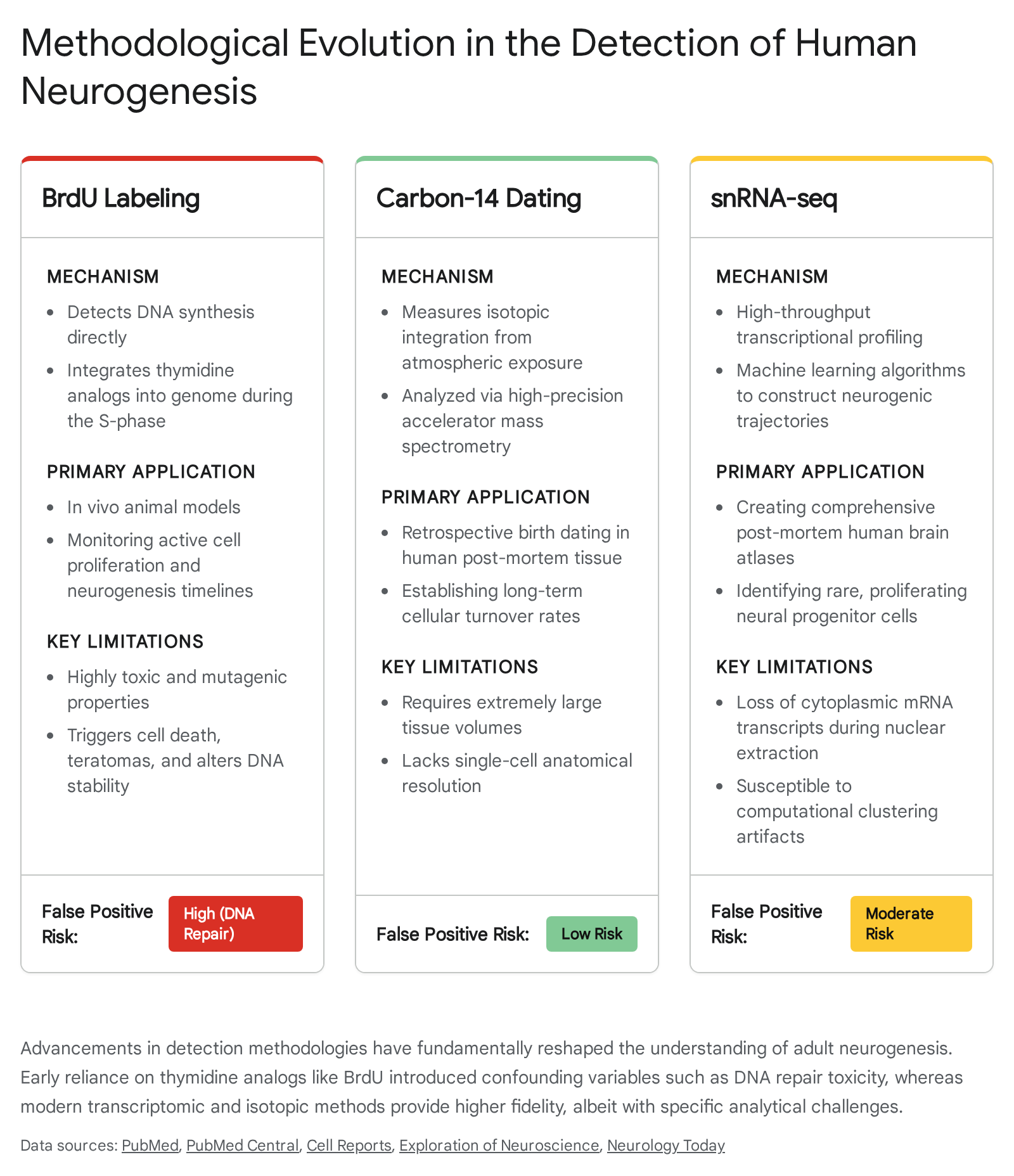
Thymidine Analogs and Bromodeoxyuridine Toxicity
For several decades, the gold standard for identifying adult-born neurons in situ was the exogenous administration of thymidine analogs, predominantly Bromodeoxyuridine (BrdU). BrdU incorporates into the newly synthesized DNA of dividing cells specifically during the S-phase of the cell cycle. When tissue is later analyzed via immunohistochemistry, BrdU-positive cells co-labeled with mature neuronal markers (such as NeuN) are traditionally interpreted as newly generated neurons 42526.
However, the application of BrdU presents severe analytical pitfalls. BrdU is not an exclusive marker of mitosis; it is fundamentally a marker of DNA synthesis. Consequently, it incorporates during various non-proliferative cellular events, including DNA repair processes, gene duplication, and abortive cell cycle re-entry 262728. This is particularly problematic in the context of neurodegenerative conditions like Alzheimer's disease, where mature, dying neurons frequently re-enter the cell cycle, replicate their DNA, and subsequently undergo apoptosis without completing mitosis. If labeled with BrdU, these degenerating neurons generate false-positive signals that mimic neurogenesis 2628. Furthermore, BrdU is intrinsically toxic and mutagenic. It alters DNA stability, triggers apoptosis, lengthens the cell cycle, and disrupts the very neurogenic processes it is designed to measure, severely restricting its ethical and practical application in living humans 272930.
Retrospective Carbon-14 Dating
To circumvent the toxicity and artifact risks of exogenous labeling, researchers developed retrospective carbon-14 ($^{14}C$) dating. This methodology leverages the dramatic spike in atmospheric $^{14}C$ concentrations caused by above-ground nuclear bomb testing between 1955 and 1963. Because atmospheric $^{14}C$ levels rapidly declined following the test ban treaty, the concentration of the isotope integrated into a cell's genomic DNA during mitosis accurately reflects the atmospheric concentration at the specific time of cell division 83132.
By utilizing fluorescence-activated cell sorting (FACS) to isolate neuronal and non-neuronal (glial) nuclei from post-mortem human brains, and analyzing their $^{14}C$ content via accelerator mass spectrometry, researchers mapped cell turnover dynamics over decades 833. This approach demonstrated that while neocortical neurons are virtually as old as the individual - indicating no substantial turnover - hippocampal dentate gyrus neurons display measurable, continuous turnover throughout the lifespan 83233. Mathematical modeling of the $^{14}C$ integration data estimated that approximately 700 new neurons are added to each human hippocampus daily. This reflects an annual turnover rate of roughly 1.75%, which declines only modestly with advanced age 83435. Crucially, investigations into background DNA repair confirm that post-mitotic repair mechanisms exchange orders of magnitude too little carbon to account for the observed $^{14}C$ concentrations, validating the method as an accurate metric of de novo cell birth rather than DNA maintenance 836.
Single-Nucleus RNA Sequencing
In recent years, single-nucleus RNA sequencing (snRNA-seq) has revolutionized the transcriptomic profiling of the brain. Unlike single-cell RNA sequencing (scRNA-seq), which requires fresh tissue and harsh enzymatic dissociation that biases against delicate, complex cells, snRNA-seq allows for the high-throughput transcriptomic profiling of isolated nuclei from frozen post-mortem human tissue 37383940.
By analyzing the transcriptome at single-nucleus resolution, researchers can identify distinct cell states and reconstruct the developmental trajectories of neurogenic lineages across the lifespan using computational tools like pseudotime mapping 2042. However, snRNA-seq involves profound analytical challenges. Because it relies entirely on nuclear RNA, it inherently misses highly abundant cytoplasmic mRNA, substantially reducing its sensitivity to detect low-expression markers 373940. The inherent stochastic noise in snRNA-seq data risks generating false positives, particularly when searching for exceptionally rare cell populations. Furthermore, discrete clustering algorithms may force continuous, dynamic cell states into artificial categories, complicating the identification of true transient progenitor states 394041.
Spatial Transcriptomics and Validation
To ground computationally derived snRNA-seq data in anatomical reality, recent studies utilize highly multiplexed spatial transcriptomics (e.g., RNAscope, Xenium in situ). These technologies allow researchers to visualize the spatial expression of hundreds of transcripts directly within the intact tissue architecture 24374243. This is an essential validation step, as standard imaging-based immunohistochemistry in human tissue is often confounded by lipofuscin autofluorescence - a ubiquitous byproduct of cellular aging that frequently generates false-positive signals 1937.
The Contemporary Debate on Human Hippocampal Neurogenesis
The application of high-resolution transcriptomic and immunohistochemical technologies has resulted in highly polarized scientific conclusions regarding the existence, volume, and persistence of adult human neurogenesis.
Evidence for Rapid Postnatal Cessation
A series of high-profile studies have argued that adult hippocampal neurogenesis in humans is an overwhelmingly juvenile phenomenon that drops to negligible or undetectable levels by early childhood or adolescence. In a landmark 2018 study, Sorrells et al. analyzed human tissue ranging from prenatal stages to senescence. They reported a precipitous decline in dividing cells and young neurons during the first year of life, concluding that neurogenesis drops to virtually undetectable levels in subjects older than 13 years 219233442.
This conclusion was reinforced by Franjic et al. (2022), who utilized a comprehensive cross-species snRNA-seq approach. While they identified robust, continuous neurogenic trajectories in the hippocampi of adult mice, pigs, and macaques, they detected no such continuum in the adult human dentate gyrus. Out of over 32,000 human granule cells sequenced, only a single cell exhibited a distinct neuroblast-like transcriptomic profile 420424445. Similarly, spatial transcriptomic analyses conducted by Simard et al. (2024) detected substantial populations of DCX-expressing cells in the human hippocampus across all ages, but failed to detect the proliferative stem cell markers necessary to prove these cells were actively dividing and differentiating. They concluded that while a local reserve of plasticity exists, continuous de novo cell birth is exceptionally rare 472342.
Evidence for Sustained Lifelong Production
Conversely, a robust body of evidence maintains that neurogenesis persists throughout the human lifespan, albeit at lower rates than observed in rodents. Boldrini et al. (2018) reported similar levels of intermediate neural progenitors and thousands of immature neurons across the lifespan into the eighth decade, maintaining that angiogenesis and neurogenesis continue in healthy aging 243448.
Further supporting this view, independent groups re-evaluated human snRNA-seq datasets using distinct computational frameworks. Using supervised machine learning algorithms rather than standard unsupervised clustering, Zhou et al. (2022) and Wang et al. (2022) successfully identified populations of immature granule cells and proliferating NSCs that previous analyses had miscategorized or overlooked due to their transcriptional similarity to mature glia and neurons 71517204448.
The most definitive recent evidence emerged in a 2025 Science publication by Dumitru, Paterlini, and Frisén. Acknowledging the extreme scarcity of proliferating cells, the researchers bypassed standard bulk sampling. Instead, they utilized flow cytometry to enrich post-mortem hippocampal tissue specifically for nuclei expressing Ki-67 (a definitive marker of active cellular proliferation). By training a machine-learning model on data from robust childhood neurogenesis, they successfully identified actively dividing neural progenitor cells and their subsequent neuroblast lineages in humans ranging from infancy up to 78 years of age 84346474849. Validated by RNAscope and Xenium spatial transcriptomics, this study confirmed the active generation of new neurons. However, the authors explicitly cautioned that while their qualitative methodology proves the existence of the process, it cannot determine the absolute quantitative frequency of these events, noting that baseline neurogenesis varies significantly among individual adults 43464748.
Protracted Maturation and Human Neoteny
The difficulty in detecting classic, rapidly dividing neurogenic lineages in humans has led to the prominent theory of protracted maturation, or neoteny. In rodents, a newborn neuron matures, extends its axons, and functionally integrates into the hippocampal circuit within 3 to 4 weeks. In contrast, neurons in primates take several months, and in humans, potentially years or decades, to fully mature 121719.
This phenomenon suggests that many of the cells expressing "immature" markers like DCX or PSA-NCAM in the adult human brain are not necessarily newly born. Instead, they may be neurons generated during late fetal or early postnatal development that have been held in a state of arrested development or prolonged immaturity 923505152. Support for this concept comes from transcriptomic analyses indicating that adult immature neuron pools exhibit "molecular ages" characterized by high levels of DNA repair, ribosomal function, and telosome (shelterin complex) maintenance. This profile suggests these cells are chronologically older than actively dividing, newly born neuroblasts, but maintain a juvenile transcriptional program to support plasticity 5053.
Parenchymal Immature Neurons Outside the Niche
This state of developmental arrest is not unique to the hippocampus. Protracted maturation is heavily documented in non-canonical regions such as the paralaminar nucleus of the human amygdala and layer II of the paleocortex and neocortex. In these regions, vast populations of developmentally stalled, DCX-positive excitatory neurons exist in a dormant state 9525754. During childhood, adolescence, and potentially adulthood, a subset of these dormant neurons gradually resumes structural differentiation, expanding their dendritic arbors and integrating into local circuits without requiring the division of a stem cell. This alternative mechanism is often described as "neurogenesis without division" or parenchymal neuroplasticity 9525754.
Cross-Species Divergence in Neuroplasticity
Transcriptomic and morphological studies consistently reveal that adult neurogenesis is not a perfectly conserved biological mechanism across all mammalian species. The evolutionary transition from small-brained, lissencephalic species (rodents) to large-brained, gyrencephalic mammals (primates, dolphins, and humans) is accompanied by significant trade-offs in neuroplastic processes 95254.
Evolutionary Trade-Offs in Brain Size
While rodents maintain a highly active NSC pool that continuously adds thousands of neurons to the olfactory bulb and hippocampus, this continuous generative capacity is vastly downgraded in larger mammals. Interestingly, species with exceptionally large brains and long lifespans, such as dolphins and whales, appear to lack adult neurogenesis almost entirely 135254. The evolutionary trade-off hypothesis posits that instead of relying on active, continuous stem cell division - which carries cumulative metabolic costs and risks of tumorigenesis - larger-brained mammals favor the early generation and subsequent preservation of dormant, immature neurons within the parenchyma 952.
Furthermore, species-specific molecular regulation governs the neurogenic niche. Single-nucleus transcriptomics of the macaque hippocampus has identified dozens of genes expressed in primate neural stem cells that are completely undetectable in the mouse dentate gyrus. For example, the enzyme ethanolamine-phosphate phospho-lyase (ETNPPL) is robustly expressed in actively proliferating primate SGZ stem cells, functioning as a primate-specific NSC marker 7151720. Similarly, STMN1 and STMN2 have emerged as novel markers of immature granule cells specifically in primates 2052. This evolutionary divergence has profound implications for translational research, indicating that mouse models may not accurately recapitulate the baseline cellular turnover rates or the exact molecular identity of human neural stem cells 4754.
| Feature / Metric | Murine (Mouse) Model | Primate & Human Model |
|---|---|---|
| Olfactory Bulb Neurogenesis | Robust, lifelong migration via SVZ | Effectively absent in adulthood |
| Hippocampal Maturation Rate | Rapid integration (3 - 4 weeks) | Protracted (months to years/decades) |
| Proliferation Volume | High (thousands of cells per day) | Exceedingly low; highly variable between individuals |
| Parenchymal Immature Neurons | Scarce (localized mainly to Piriform cortex) | Abundant (Paralaminar amygdala, Neocortical Layer II) |
| Specific NSC/imGC Markers | Sox2, GFAP, Nestin, Prox1 | ETNPPL (Primate specific NSC), STMN1/2, Sox2, GFAP |
Modulators of Adult Hippocampal Neurogenesis
Despite the low baseline rate of cell division in humans, the hippocampal neurogenic niche remains highly responsive to both internal physiological states and external environmental stimuli. The modulation of this niche occurs at multiple levels, influencing stem cell activation, neuroblast proliferation, and the survival of integrating immature neurons.
The Dorsoventral Functional Segregation
The modulation of neurogenesis is anatomically segregated along the dorsoventral axis (analogous to the posterior-anterior axis in primates). The dorsal hippocampus is primarily integrated into cortical networks governing spatial navigation, explicit learning, and cognitive flexibility. Baseline neurogenesis is naturally higher in this dorsal region, and it is preferentially upregulated by environmental enrichment and physical exercise 555657.
Conversely, the ventral hippocampus is heavily interconnected with the amygdala and the hypothalamic-pituitary-adrenal (HPA) axis, regions governing emotional regulation, anxiety, and the physiological stress response. Neurogenesis in the ventral region exhibits greater overall plasticity but is highly susceptible to the suppressive effects of chronic stress, indicating that the local microenvironment dictates how neurogenic output responds to systemic signals 555657.
Extrinsic Modulators: Exercise and Diet
Aerobic physical activity is established as one of the most potent positive modulators of adult hippocampal neurogenesis across species. Voluntary exercise upregulates the peripheral production of metabolic and vascular growth factors - most notably Brain-Derived Neurotrophic Factor (BDNF), Insulin-like Growth Factor 1 (IGF-1), and Vascular Endothelial Growth Factor (VEGF). These factors cross the blood-brain barrier to promote angiogenesis, NSC proliferation, and the synaptic integration of adult-born neurons 105662. Environmental enrichment (EE) - exposure to complex, stimulating environments - primarily acts to enhance the functional integration, dendritic complexity, and long-term survival of immature neurons rather than strictly driving precursor proliferation 11062.
Dietary interventions also exert profound structural effects. Caloric restriction, intermittent fasting, and the intake of specific neuroprotective polyphenols (e.g., curcumin, cocoa flavanols) and omega-3 fatty acids stimulate neurogenic activity by lowering systemic oxidative stress and upregulating neurotrophic signaling cascades. In some animal models, meal frequency and fasting intervals have been shown to be more critical than absolute caloric restriction in promoting NSC activation 535556258. Conversely, high-fat, high-sugar diets and early-life malnutrition drastically impair NSC proliferation by inducing chronic neuroinflammation and disrupting metabolic homeostasis within the niche 56258.
Intrinsic Modulators: Aging, Stress, and Hormones
Chronological aging is the primary intrinsic negative modifier of neurogenesis. As organisms age, the local microenvironment grows less permissive to neurogenesis due to increased baseline inflammation and the accumulation of senescent cells, pushing the neural stem cell pool into a deep, quiescent dormancy 315.
Chronic stress exerts a similarly deleterious effect on the neurogenic niche. Prolonged exposure to elevated glucocorticoids (cortisol in humans, corticosterone in rodents) heavily suppresses cell proliferation in the ventral dentate gyrus. Stress-induced neuroinflammation involves the elevation of pro-inflammatory cytokines such as IL-1β, IL-6, and TNF-α. These cytokines actively disrupt NSC differentiation, impair structural maturation, and force immature cells into apoptotic cell death pathways, linking chronic stress directly to hippocampal atrophy 3105564.
Hormonal status further dictates neurogenic output. Estrogens, primarily acting through receptors such as ERβ and GPER1 in the dentate gyrus, stimulate cell proliferation and modulate the survival of new neurons in a dose-dependent manner. Similarly, Erythropoietin (EPO), traditionally recognized for its hematopoietic role, has been shown to enhance neurogenesis and inhibit the apoptosis of newborn cells following cognitive challenge or hypoxia 5659.
Neurogenesis in Pathological Contexts
The impairment or alteration of adult neurogenesis is increasingly recognized as a contributing factor in the pathogenesis of various neuropsychiatric and neurodegenerative diseases, making it a prime target for therapeutic intervention.
Reactive Neurogenesis Following Brain Injury
Under healthy physiological conditions, canonical neurogenesis is restricted to the SVZ and SGZ, with the human neocortex remaining entirely non-neurogenic 146061. However, severe neurological insults, such as ischemic stroke or traumatic brain injury (TBI), drastically alter the local biochemical environment. Extensive cell death triggers the release of damage-associated molecular patterns and potent growth factors (such as EGF and FGF-2), which can coax endogenous neural precursors from the SVZ to proliferate and migrate toward the site of injury in the striatum or neocortex 22606263.
In animal models, transient populations of neuroblasts have been observed migrating toward damaged tissue following ischemia. Unfortunately, these non-canonical, damaged environments lack the highly specialized extracellular matrix, vascular support, and specific trophic cues of a true neurogenic niche. Consequently, the vast majority of these recruited cells fail to terminally differentiate, form appropriate synaptic connections, or survive long-term. Thus, while severe injury induces an initial, reactive neurogenic response, it does not currently lead to meaningful structural or functional self-repair in the adult human neocortex 142262.
Neurodegenerative and Psychiatric Implications
The physiological purpose of adult-born dentate granule cells lies in their unique electrophysiological properties during their protracted immature phase. Their hyper-excitability allows them to act as highly sensitive pattern separators. By incorporating new, highly plastic neurons into the CA3 network, the hippocampus can encode novel information with minimal interference to previously established circuits 7151621.
When this process is disrupted, cognitive and emotional regulation falter. In major depressive disorder, the suppression of neurogenesis via chronic stress pathways correlates strongly with hippocampal volumetric decline and emotional dysregulation. Crucially, the behavioral efficacy of many standard antidepressant medications appears to rely, at least in part, on their ability to upregulate adult hippocampal neurogenesis and restore structural plasticity to the ventral hippocampus 35558.
In Alzheimer's disease, a sharp decline in the molecular signatures of immature neurons correlates directly with the progression of dementia. Transcriptomic profiling of brain tissue from individuals who exhibit advanced Alzheimer's pathology (amyloid plaques and tau tangles) but remain cognitively intact - a phenomenon known as cognitive resilience - reveals a remarkable preservation of immature neuronal networks. These networks actively engage in neurotrophic, anti-inflammatory, and anti-amyloidogenic signaling pathways, suggesting that a robust reserve of immature neurons may actively buffer the brain against neurodegeneration and maintain cognitive homeostasis 9485053.
Moving forward, the field of regenerative medicine seeks to harness this endogenous plasticity. The definitive identification of proliferating progenitors in the adult human brain up to the eighth decade provides foundational proof-of-concept that the human brain retains a latent regenerative capacity 84349. Future therapeutic interventions will likely shift focus from simply forcing dormant stem cells to divide, toward modulating the inflammatory and metabolic microenvironment of the aging niche. By creating a permissive environment, therapies may eventually support the protracted survival, maturation, and functional integration of adult-born neurons to combat cognitive decline 3384862.